Download
1 / 48
800 likes | 1.93k Views
ASSESSMENT OF THE ABDOMEN. Prepared by Hamdia Mohammed. Learning Objectives:-. At the end of this lecture each student will be able to: Identify landmarks for the abdominal assessment Correctly perform techniques of inspection, auscultation, percussion and palpation

E N D
ASSESSMENT OF THE ABDOMEN Prepared by Hamdia Mohammed
Learning Objectives:- At the end of this lecture each student will be able to: • Identify landmarks for the abdominal assessment • Correctly perform techniques of inspection, auscultation, percussion and palpation • Differentiate between normal & abnormal findings.
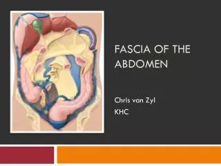
Overview of abdominal structure. 1- Large oval cavity. 2- Extends from diaphragm to symphysis. 3- Viscera: solid and hollow. A- Solid viscera are those organs that maintain their shape consistently ( liver, pancreas, spleen, adrenal glands, kidneys, ovaries and uterus ).
The liver is the largest solid organ in the body. B- The hollow viscera consist of structures that change shape, depending on their contents . These include ( stomach, gallbladder, small intestine, colon , bladder ). 4- Vascular structures: The abdominal organs are supplied with arterial blood by abdominal aorta & its major branches.
Locating abdominal structures by quadrants Divided to four quadrants:- 1- Right upper quadrant ( RUQ ) 2- Right lower quadrant ( RLQ ) 3- Left upper quadrant ( LUQ ) 4- Left lower quadrant ( LLQ )
Right upper quadrant ( RUQ ). Left Upper Quadrant (LUQ ). - stomach - spleen - left lobe of liver - body of pancrea - left kidney and adrenal - spleen flexure of colon - part of transverse & descending colon - Liver - Gallbladder - Duodenum - Head of pancreas -Right kidney and adrenal - Hepatic flexure of colon - Part of ascending and transverse colon. - Right ureter.
Right Lower Quadrant: Left Lower Quadrant: -Part of descending colon -Sigmoid colon -Left ovary and tube -Left ureter -Left spermatic cord -Cecum -Appendix -Right ovary and tube -Right ureter -Right spermatic cord Midline: -Aorta -Uterus. -bladder.
Preparation for abdominal assessment • Preparing the exam room • preparing the patient • positioning the examiner
Health History: • Any chronic diseases that affect GIT or urinary systems? Describe. • Does he drink alcohol? How much? How often? When was last drink? • Smoke? How much and how long? Considered stopping or cutting down? • How often do you have a bowel movement? When was the last one? What are color and consistency of stool?
Nausea or vomiting for how long? Frequency? • How much do vomit? What does it look like? Contain blood? Have an odor? • Abdominal pain: • How long have he had ? Where? When did he first feel pain? What activity were he doing? • Describe pain. Constant/intermittent? Had episodes before? Did pain start suddenly?
Types of pain • Vesceral pain. • Parietal pain : as in appendicitis • Referred pain
Character of abdominal pain • Dull, aching( e.g cystitis ) • Burning (e.g dyspepsia ) • Colicky (e.g colon cancer) • Sharp, knifelike (e.g renal colic ) • Pressure ( urinary retention )
Assessment Techniques 1- Inspection. 2- Auscultation. 3- Percussion. 4- Palpation.
1- Inspection • skin: color, scars, veins, lesions. • umbilical hernia, bleeding, inflammation. • contour of the abdomen :flat ,rounded, protuberant . • symmetry • enlarged organ. • Masses. • Peristalsis ,pulsation , distention.
Distention:- Definition: unusual stretching of abdominal wall Abdominal distention can be caused by three factors: 1. Obesity – Abdomen is soft and rounded with a sunken umbilicus. 2. Ascites – Skin is shiny and glistening with an everted umbilicus. Veins are dilated and prominent (more visible in thin, malnourished skin). 3. Obstruction – There may be visible, marked peristalsis; restlessness; lying with knees flexed; grimacing facial expression; and uneven respirations.
Distention:- • note position of umbilicus • note portion of abdomen that is distended • reasons for distention: flat(obesity), flatus(gas), feces, fluid, tumor , fetus(pregnancy )
2- Auscultation • Auscultation performed before palpation and percussion. • Use diaphragm of stethoscope • Listen to bowel sounds for up 5 minutes in each quadrant. • Normal sounds are clicks and gurgles, irregular, 5-30 times per minute • Influenced by digestion
Auscultation con’t • Increased bowel sounds are due to hypermotility of peristalsis • Decreased are due to paralytic ileus or peritonitis • Intestinal obstruction can present with increased or decreased sounds
Abdominal Vessels Sites for Auscultating the Abdomen
Additional Sounds Bruits: • Bruits are low pitched, vascular sounds, resembling murmur • Caused by partially obstructed artery– turbulence • Listen in epigastrum and each upper quadrant • Listen in costovertebral angle(with patient seated) • Listen over aorta, iliac arteries, femoral arteries • Arterial insufficiency in legs
3- Percussion • Assessment technique used to assess size and density of organs in the abdomen e.g used to measure size of liver or spleen. • In the right midclavicular line, percuss down from lung resonance to liver dullness.
Percussion con’t • Used to identify air in stomach or in bowel. • Used to identify masses. • Used alone or in conjunction with palpation or to validate palpatory findings. • Orient to the abdomen by lightly percussing all 4 quadrants for tympany or dullness.
Percussion con’t • Tympany usually predominates due to gas in the bowel. • Dullness may be present due to feces or fluid or over organs or a solid mass. • Develop a specific percussion route and stick to it.
Percussing the spleen • Where is the spleen located? In the curve of the diaphragm just posterior to the left midaxillary line. • When the spleen enlarges, it does so anteriorly, downward and medially. This will replace the tympany of the stomach and colon with dullness
Tricks to Assessing the Spleen • Percuss in the lowest interspace in the left anterior axillary line for tympany. • Ask the patient to take a deep breath and percuss on inspiration. • The percussion note should remain tympanic. • A change to dullness suggest spenomegally • This is known as a positive splenic percussion sign
Percussion Sites for all Quadtrants (Abdominal percussion seqences may proceed clockwise)
4- Palpation • To differentiate voluntary from involuntary resistance: rectus muscle will relax with expiration. • Palpation is light or deep • Deep palpation used to define and delineate organs or abdominal masses. • Use palmar surface of fingers and feel in all four quadrants
Palpation con’t • Used to assess muscle tone, tenderness, fluid, organs. • Use pads of fingertips in light dipping motions and avoid short jabs.
Palpation of the liver • Stand on patients right side • Place left hand behind patient parallel to and supporting 11-12th ribs • Patient should relax • Press with left hand forward and place right hand on abdomen with fingertips below lower edge liver dullness • Press in and up while patient takes deep breath; if palpable, liver should come down
Palpation of the spleen • The spleen is usually not palpable • From patient’s right side, reach over and around under patient with left hand • Place right hand below left costal margin and press in toward spleen. Ask patient to take deep breath---will feel if palpable
B- Auscultating the abdomen A-Inspecting the abdomen D- Percussing the abdomen C- Palpating the abdomen
Learning Objectives:- 1- Identify the important new terms related to urinary system. 2- List the factors which influencing urination. 3- Enumerate function of kidneys. 4- Differentiate between normal and abnormal finding.
Important new Terms * Oliguria: voiding a scanty amount of urine. * Anuria:inability to produce urine, less common, but caused by a decrease in renal perfusion. * Polyuria: excessive output of urine. * Hematuria: blood noted in urine. * Nocturia: having to void at night.
* Dysuria: difficulty in voiding or pain in voiding. * Enuresis: involuntary loss of urine at night. * Pyuria : presence of pus in the urine. * Glycosuria: presence of sugar in urine. * Albumin urea: presence of albumin in the urine.
Factors influencing urination Socio cultural psychological muscle tone fluid balance surgical procedures medication
Functions of the kidneys Kidney:- •Urine formation • Excretion of waste products • Regulation of electrolytes • Regulation of acid–base balance • Control of water balance • Control of blood pressure • Renal clearance • Regulation of red blood cell production • Synthesis of vitamin D to active form • Secretion of prostaglandins.
Palpation of kidney • Find the costovertebral angle which formed by the lower border of the 12th rib and the transverve processes of the upper lumbar vertebrae. • Place left hand flat in this area on one side, hit the hand sharply with the fist of the other patient will admit to tenderness if present. • Repeat on the other side
Palpation cont. • Kidney: not palpable in normal adult. • May be able to feel lower right kidney pole in very thin person. Technique for palpating the right kidney (top). Technique for palpating the left kidney.
Deep palpation • If masses are felt, note: location, size, shalpe, consistency, tenderness, pulsations, mobility with respiration or with hand. • If patient is obese or rigid, use 2 hands to palpate • Place one on top of other and feel with lower hand
Palpation of the bladder • Bladder percussion is unnecessary unless there is a suspicion of urinary retention. • Palpate above the symphysis. • An empty bladder is not palpable.
The bladder should be percussed after the patient voids to check for residual urine. • Percussion of the bladder begins at the midline just above the umbilicus and proceeds downward. • The sound changes from tympanic to dull when percussing over the bladder.
The bladder, which can be palpated only if it is moderately distended, feels like a smooth, firm, round mass rising out of the abdomen, usually at midline. • Dullness to percussion of the bladder following voiding indicates incomplete bladder emptying.
Assessing the Aorta • Press firmly deep in upper abdomen slightly to left of midline. • Feel for aortic pulsations • Determine width of aorta by pressing deeply on either side of aorta • What is the normal width of the aorta? • If pulsatile mass is found, feel for femoral pulses which may be dimished.
Special test for appendicitis: Rebound tenderness: mean deeply palpation& withdrawal quickly, this caused pain in appendicitis. Psoas sign : pt lie in supine position & raise right leg , if the pain found this is indicate to appendicitis. Oburator sign: pt flex right leg at hip and knee. Then rotate leg internally and externally.