Inflammatory Process
Inflammatory Process. Inflammation. What is Inflammation A vascular and cellular response to trauma. Its purpose is to initiate the healing of the injured tissue The body’s attempt to dispose of micro-organisms, foreign material and dying tissues so that tissue repair can occur

Inflammatory Process
E N D
Presentation Transcript
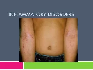
Inflammation • What is Inflammation • A vascular and cellular response to trauma. Its purpose is to initiate the healing of the injured tissue • The body’s attempt to dispose of micro-organisms, foreign material and dying tissues so that tissue repair can occur • An inflammatory response may result from external or internal factors (infection) • Protects to the body by localizing and removing the injuring agent
Signs of Swelling • Redness (Rubor) • Swelling (Tumor) • Pain (Bolar) • Warmth (Calor) • Loss ROM
Signs of Inflammation (Cardinal Signs) • Redness (Rubor): • Caused by blood vessel dilation (the arterioles) • Chemical mediators promote the vessel dilation (contained in the capillary walls or endothelium resulting in immediate response) • Histamine • Seritonin • Bradykinins • Prostaglandins • Note: a 1x increase in arteriole diameter yields a 4x increase in blood flow
Signs of Inflammation Cont. • Swelling (tumor) • Edema fluid varies with the stage of inflammation • initially vessel permeability is only slightly altered and no cells or protein escapes and the fluid is mainly water and dissolved electrolytes (transudate): like synovial fluid • As capillary permeability increases and plasma proteins escape the extravascular fluid becomes cloudy and more viscous. This is called exudate (contains a large amount of leukocytes (called pus)
Causes of Edema/Swelling- • bleeding from torn vessels • cell death due to anoxia, allows fluid leakage (permeability increases) • increased proteins raise extracellular osmotic pressure, drawing fluids from the capillaries • Chemicals alter cell permeability to proteins and fluid • Gravity may increase swelling (Capillary filtration pressures)
Edema/Swelling • To cease hemorrhage/swelling/edema • Must reverse the condition • pressure gradient • vessel repair • This is what we try to do as therapists through modality use
Signs of Inflammation Cont. • Pain (bolar) • Results from irritation of nerve ending by physical or chemical factors • Physical trauma may irritate pain receptors • Chemical mediators release when cell damage occurs sensitize pain receptors • Trauma may result in cell anoxia because of interference with blood flow due to capillary damage
Signs of Inflammation Cont. • Warmth (calor) • The result of chemical activity and increased blood flow in the injured area. • Loss of Function • May occur due to pain causing reflex guarding or muscle spasm, spasm decreases metabolic activity and constricts blood flow which causes more pain due to ischemia; thus the pain cycle
Phases of the Inflammatory Process • Phase I: Acute Phase • inflammatory response: lasts 2-4 days but is complete in 2 weeks • Phase 2: Tissue Formation (Proliferation) • Subacute phase, Tissue rebuilding approximately 2-3 weeks • This does not include chronic inflammation • Phase 3: Remodeling Phase • Adapt to original tissue • Continues for up to 1 year post injury
Phase I: The Inflammatory Process • Early Phase • Insult occurs - may be internal (infection) or external (trauma) • Vasoconstriction to decrease blood flow (first 10 minutes) • Vasodilatation • Late Phase • Tissue Repair • Regeneration
Inflammatory Phases Chart Designates Percent of phase over time
Phase I: Early Phase Inflammation - Vasodilatation • Chemical mediators are released: • histamine, bradykinis, serotonin, prostaglandin's - increase vascular permeability released from mast cells and blood platelets into traumatized tissue. • As fluid filtrates through “gaps in the extravascular spaces this is calls exudation.
Phase I: Vasodilatation Cont. • The accumulation of excess fluid is called edema (Swelling) • Vascular permeability due to action of the histamine is short-lived, lasting less than 1 hour
Phase I: Early Phase Inflam. - Lymphatic channels are blocked • Local lymphatic channels are blocked by fibrin plugs formed during coagulation. Obstruction of the local lymphatic channels prevents drainage of fluid from the injured site, thus localizing the inflammatory reaction.
Phase I: Early Phase Inflammation - Margination • When trauma occurs the endothelial wall is disrupted exposing collagen fibers creating a “stickiness” • WBC’s concentrate in the injury site to rid the body of foreign substances and dead (necrotic) tissue
Phase I: Margination Cont. • As circulation slows, leukocytes migrate and adhere to the walls of post-capillary venuels (for approx 1 hour) • The leukocytes pass through the walls of the vessels (diapedesis) and travel to the site of injury (Chemotaxis)
Phase I: Early Phase Inflammation - Phagocytosis • Body’s cellular defense to remove toxic material via lymphatic system • Phagocytosis: a process when leukocytes capture and digest foreign matter and dead tissue • 1st line of defense: neutrophiles(in most abundance from 1-3 days) - phagocytic activity reaches maximum effectiveness within 7-12 hours
Phase I: Phagocytosis Cont. • 2nd line of defense: monocytes (which convert into large cells called macrophages) and lymphoctes consume large amounts of bacteria and cellular debris. Monocytes are critical in the initiation of tissue repair because the attract fibroblasts Bacteria Macrophage
Phagocytosis Cont. • Pus is the end result - it contains leukocytes, dead tissue and phagogenic material • Prolonged puss accumulation can prevent fibroplasia which begins the wound healing • Fibrobalsts are connective tissue responsible for collagen synthesis • Ligaments, joint capsule, tendon • Osteoblasts: responsible for bone synthesis Fibropblast Macrophages
Phase I: Late Phase Blood Clotting • Ruptured vessels release Enzyme (Factor X) • Factor X reacts with prothrombin (free floating in blood) • Thrombin then stimulates fibrogen into its individual form fibrin • Fibrin grouped together to form “lattice” around injured area • Fibrin lattice contracts to remove plasma and compress platelets forming a “patch”
Phase I: Late Phase Blood Clotting Factor X Prothrombin Fibrin Forms Seal Thrombin Fibrin Mesh Fibrogen and Thrombin Meet Fibrin Monomer
Phase II: Regeneration: • The replacement of destroyed cells by reproducing healthy cells adjacent to the wound (humans capacity to regenerate tissue is limited and further affected by age and nutritional state).
Phase II: Stages of Regeneration: • Stage starts with periphery • Re-eptheliaization is proliferation of peripheral epithelial tissue which then migrates to the wound until the area is covered. • Capillarization (Capillary buds proliferate and connect forming new capillaries which gives the red, granular appearance to the scar (granular tissue)
Phase II: Stages of Regeneration: Cont. • Fibroplasia occurs due to fibroblasts which arises from undifferentiated mesenchymal cells and migrate into the area along fibrin strands and begin to synthesize scar tissue. Scar tissue is CT and mostly collagen and mucopolysaccharides. Fibroblasts secrete both, contributing tensile strength to the repair. Scar tissue very inelastic compared to surrounding tissue.
Phase II: Stages of Regeneration: Cont. • Vascularization - occurs with the proliferation of collagen synthesis • Formation of blood vessels (angiogensis)
Phase II: Collagen Synthesis: • Occurs within 12 hours of injury to 6 weeks (average 3 weeks) • Type I: collagen: associate with muscular tissue (larger and stronger fibers) • Type III collage: smaller fibers, less cross linking and highly disorganized (ligamentous, tendinous) • Type III with time is replaced by Type I collagen
Phase II: Collagen Synthesis Cont. • Tissue Healing Times • Muscle : approximately 3 weeks • Tendon: 4-6 weeks • Extent of the tissue damage and vascularity will aid in determining healing time • Age may also be a factor in healing
Phase II: Stages of Regeneration: Cont. • Wound Contraction: • Wound contraction begins to occur in CT as the myobroblasts (actin-rich fibroblasts) contract. Myofibroblasts move toward the center of the wound, helping reduce the size of the area to be covered. • Outside-in
Phase III: Maturation/Remodeling Phase • Purpose of this phase • Strengthen the repaired tissue • Firoblasts, myofobrpb;asts & Macrophages reduced to preinjury state • Type I fibrin continues to be replaced by type III
Phase III: Maturation/Remodeling Phase (day 9 onward) • Blends in with the repair phase, original collagen fibers were randomly oriented. During remodeling, the fibers become more organized, parallel to the wound surface which provides greater tensile strength • The type of tissue involved will determine the duration and extent of remodeling activity
Phase III: Maturation/Remodeling Phase Cont. • Strengthening of scar tissue continues from 3 months to 1 year, but fully mature scar in only 70% as strong as intact tissue. • Motion will influence the structure and functional capacity of scar tissue (controlled stress increases functional capacity, allows healing and reduces adhesion formation).
Chronic Inflammation • Inflammation which continues past 1 month • Marked by a loss of function • Fibroblast activity continues forming granuloma
Chronic Inflammation • Complications • Granuloma: large mass of weaker scar tissue (usually due to large inflammation and activity without regard to healing time) • Retardation of muscle fiber: with excessive granuloma fibroblasts cannot reach damaged tissue • Adhesions/contractures in tissue • Keloid/hypotrophic scars
Abnormal scarring: • Hypertophic scar or keloid scar. Biological difference not well understood, but clinically hypertrophic scar is contained within the boundaries of the original wound while a keloid scar extends beyond the borders of the original wound.
THE BIG QUESTIONS! • When do we use cold? • When do we use heat? • When do we use medications? • When do we use Electrical modalities?