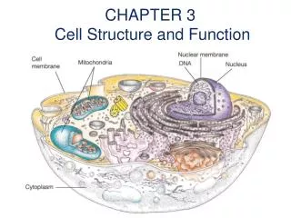
Understanding Human Chromosome Structure & Function
Explore the intricate organization and functions of human chromosomes, including DNA packaging, centromeres, telomeres, and genetic diversity through meiosis and mitosis. Learn about chromosome staining techniques and karyotyping in cytogenetics.

Understanding Human Chromosome Structure & Function
E N D
Presentation Transcript
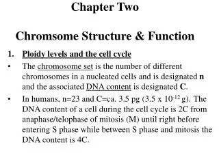
Chapter TwoChromsome Structure & Function • Ploidy levels and the cell cycle • The chromosome set is the number of different chromosomes in a nucleated cells and is designated n and the associated DNA content is designated C. • In humans, n=23 and C=ca. 3.5 pg (3.5 x 10⁻¹² g).The DNA content of a cell during the cell cycle is 2C from anaphase/telophase of mitosis (M) until right before entering S phase while between S phase and mitosis the DNA content is 4C.
In the germline, the DNA content is 2C before S phase then 4C during S until the anaphase/telophase of meiosis I. At the end of meiosis I, each reduced cell has 2C DNA content. By the end of meiosis II, each haploid gamete (egg or sperm is C).
2. Chrosmosome structure and function: • In human chromosomes, DNA is packaged in multiple hierarchies of DNA folding. • DNA wraps around a histone octamer to form the 10 nm string of beads. The latter further coil to form the 30 nm chromatin fiber (interphase). The 30 nm fibers compacts further forming a rosette shape and attaches to a scaffold of acidic proteins (topoisomersaes II). • The packaging ratio for DNA in human chromosomes is: 1:6 for nucleosomes, 1:36 for the 30 nm fiber, and >1:10,000 for the metaphase chromosome.
Chromosome organization in a human interphase is not random. Besides the nucleolus (site of rRNA synthesis) attaching to the nucleolar Organization Region (NOR), chromosomes occupy certain chromosome territories. For example, the most gene-rich chromosomes tend to concentrate at the center of the nucleus whereas the more gene-poor chromosomes tend to locate towards the nuclear envelope.
Centromere: is the primary constriction in the chromosome and the site at which the the large multiprotein complexes (kinetochore) attach to each of the centromere at late prophase. • In yeast, CEN (centromere element) is 110 bp long containing two highly conserved elements (9 and 11 bp) flanking a central AT rich region. • In mammals, centromeres consist of 100s of kilobases of repetitive DNA (some is chromosome specific). In humans, alpha-satellite is a complex family of tandemly repeated 171 bp core sequence. Several proteins such as CANP-B bind to the alpha satellite region.
Origins of replication: In simple eukaryotes (yeast) it is known as autonomously replicating sequences (ARS). In mammals, due to the lack of a genetic assay, origins of repliaction are less well defined. Mammalian artificial chromosomes seem to work without specific ARS sequences.
Telomeres: function in • Maintaining structural integrity by preventing the fusion of chromosome ends or protect the ends of chromosomes from degradation by binding to specific telomere-binding proteins. • Ensure complete DNA replication at the tips of chromosmes. • Chromosome positioning.
In a human telomere, the hexanucleotide TTAGGG is repeated to span about 3-20 kb and upstream of this repeat and heading towards the centromere exist 100-300 kb of telomere-associated repeats before any unique sequence occurs. • Telomearse is an RNA-protein enzyme that extends the 3’ overhang at the end of chromosomes.
Heterochromatin (darkly stained) and euchromatin (lightly stained): • Genes is euchromatin may or may not be expressed while genes in heterochromatin are unlikely to be expressed. • Constitutive heterochromatin. • Facultative heterochromatin (e.g. X-inactivation in females or X & Y silencing during male meiosis for a period of 15 days).
Mitosis and meiosis: • Mitosis is the somatic cell division. • Meiosis produces sperms and eggs cells
Meiosis contributes to genetic diversity in two ways: • by the independent assortment of maternal and paternal chromosomes during anaphase I. There are 223 or 8.4 million possible different combinations of parental chromosomes to be produced by one person per gamete. • Recombination (crossing over between paternal and maternal chromosomes in prophase I).
X-Y pairing and the pseudoautosomal region: • The pairing of X & Y in males is made possible by a 2.6 Mb region at the tips of their short arms. This region is known as the major pseudoautosomal region and genes in this area are not subject to inactivation and they obligatory crossover resembling normal autosomal genes (i.e. do not follow the X-linked mode of inheritance). • A 320 Kb region of homolgy between X & Y exist at the tip of their long arms but pairing and crossing over is not obligatory.
4. Visualizing human chromosomes (Cytogenetics): • Mitotic chromosomes could be stained and visualized but meiotic chromosomes are hard to visualize. White blood cells are cultured in a medium with phytohemagglutinin to induce cell division, synchronized by adding a thymidine analog, and colcemid to disrupt spindle fibre formation. • Karyotyping and chromosome banding. 46,XX or 46,XY. Human chromosomes have a total of 850 bands at high resolution. G bands (dark condensed chromatin) replicate in late S phase while G bands (light less condensed chromatin) replicate in early S phase. G bands have lower %GC content than R bands.
Molecular cytogenetics using FISH (fluorescence in situ hybridization). Probe is labeled by incorporation of fluorescent-labeled nucleotide precursors or by incorporation of a nucleotide carrying a reporter molecule (biotin or digoxigenin) which is detected that is detected by binding to a fluorescently labeled affinity molecule. • Resolution of metaphase FISH is several Mb while that of prometaphase FISH is 1 MB. For higher resolution interphase FISH is used.
Chromosome painting is achieved by using probes composed of a large collection of different DNA fragments from a single chromosome (such probes are obtained from chromosome-specific DNA libraries or by using Alu-PCR of DNA extracted from monochromosomal hybrid cells ). This technique is used to identify chromosomal rearrangements in cancer patients. • Molecular karyotyping using mixed fluorophores (one per chromosome) also known as multiplex FISH (M-FISH) allowed all 24 human chromosomes to be identified by color.
5. Chromosome abnormalities: • Result from missrepair of broken chromosomes, by improper recombination, or by malsegregation of chromosomes during mitosis or meiosis. • Two types: • Constitutional where all cells of the body have the abnormality. This results from a defective gamete or abnormal fertilization. • Somatic: occur only in certain cells or tissues of the body. This results in a mosaic individual.
Numerical chromosome abnormalities: loss or gain of complete chromosomes. - Polyploidy: where two sperms fertilize one egg (dispermy) or a diploid gamete resulting in a triploid 3n (lethal). Tetrapoild (4n) is extremely rare and results from failure to complete the first zygotic division.
Aneuploidy: one or more chromosome is missing or present in an extra copy or two. Trisomy (2n + 1) e.g. trisomy 21 (47,XX,+21 or 47,XY,+21) also known as Down Syndrome; Klinefelter (47, XXY) Monosomy (2n-1) e.g. Turner (45,X). Causes of aneuploidy are: 1. nondisjunction 2. anaphase lag.
Mixploidy: two or more genetically different cell lineages within one individual. Genetically different cell populations can arise from one zygote (mosaicism) or more rarely can originate from different zygotes (chimerism) - Aneuploidy mosaics (e.g. 2n/2n+1 are common) due to non-disjunction or chromosome lag in early mitotic divisions of the zygote. - Polyploidy mosaics (e.g. 2n/3n are occasionally found) mostly arise by fusion of the second polar body with one of the cleavage nuclei of a normal diploid zygote.
Clinical consequences of numerical abnormalities: (Table 2.4) - Autosomal monosomies are more devastating than trisomics. Trisomic embryos survive longer than monosomic ones. - sex chromosome aneuploids is less devastating than in autosomal aneupoilds. This is because of X-inactivation mechanisms and the fact that Y carries very few genes that determine male sex.
Structural chromosome aberrations: - Result from misrepair or or mal-recombination. - chromatid breaks (occur after replication in S phase) . - chromosome breaks occur in G1 phase. - misrepair of breaks could result in acentric chromosomes, dicentric chromosomes, isochromosomes. - structural abnormalities are balanced (no net gain or loss of genes) or unbalanced (there is net gain or loss of genes).
Robertsonian translocation are considered balanced even though some material is lost (the lost material is highly redundant rRNA genes). • Unbalanced abnormalities can arise through deletion, duplication, or malsegregation of chromosomes during meiosis of a balanced abnormality.