Download
1 / 26
270 likes | 604 Views
Electrical Stimulation Techniques. Current Flow. Electron Flow (shown in red) Between the generators and electrodes To and from the generator Ion Flow (shown in yellow) Occurs within the tissues Negative ions flow towards the anode and away from the cathode

E N D
Current Flow • Electron Flow (shown in red) • Between the generators and electrodes • To and from the generator • Ion Flow (shown in yellow) • Occurs within the tissues • Negative ions flow towards the anode and away from the cathode • Positive ions flow towards the cathode and away from the anode + - + -
Electrodes • Purpose • Completes the circuit between the generator and body • Interface between electron and ion flow • Primary site of resistance to current • Materials • Metallic (uses sponges) • Silver • Carbon rubber • Self-adhesive
Electrode Size • Determines the Current Density • Equal size • Bipolar arrangement • Approximately equal effects under exach
Electrode Arrangements • Based on:Current DensityProximity to Each OtherAnatomic Location (Stimulation Points)
Current Density • Bipolar Technique • Equal current densities • Equal effects under each electrode(all other factors being equal) • Monopolar Technique • Unequal current densities • At least 4:1 difference • Effects are concentrated under the smaller electrode • “Active” electrode(s) • No effects under larger electrode • “Dispersive” electrode • Quadripolar Technique • Two bipolar electrode arrangements • Two independent electrical channels • TENS is a common example “Active” “Dispersive”
Electrode Proximity • Determines the number of parallel paths • The farther apart the electrodes the more parallel paths are formed • More current is required to produce effects as the number of paths increases
Stimulation Points • Motor Points • Superficial location of motor nerve • Predictably located • Motor nerve charts • Trigger Points • Localized, hypersensitive muscle spasm • Trigger referred pain • Arise secondary to pathology • Acupuncture Points • Areas of skin having decreased electrical resistance • May result in pain reduction • Traumatized Areas • Decreased electrical resistance (increased current flow)
Path of Least Resistance • Ion flow will follow the path of least resistance • Nerves • Blood vessels • The current usually does not flow from electrode-to-electrode (the shortest path) • The path of least resistance is not necessarily the shortest path
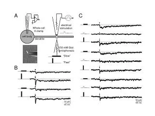
Selective Stimulation of Nerves • Nerves always depolarize in the same order • Sensory nerves • Motor nerves • Pain nerves • Muscle fiber • Based on the cross-sectional diameter • Large-diameter nerves depolarize first • Location of the nerve • Superficial nerves depolarize first
Phase Duration and Nerve Depolarization • Phase duration selectively depolarizes tissues Phase Duration Tissue Short Sensory nerves Medium Motor nerves Long Pain nerves DC Muscle fiber
Adaptations • Patients “get used” to the treatment • More intense output needed • Habituation • Central nervous system • Brain filters out nonmeaningful, repetitive information • Accommodation • Peripheral nervous system • Depolarization threshold increases • Preventing Adaptation • Vary output (output modulation) to prevent • The longer the current is flowing, the more the current must be modulated.
Electrical Stimulation Goals Muscle Contractions [Instructor Note: More detail on these techniques are found in the CH 13 ppt: Treatment Strategies]
Voluntary Type I fibers recruited first Asynchronous Decreases fatigue GTO protect muscles Electrically-induced Type II fibers recruited first Synchronous recruitment Based on PPS GTOs do not limit contraction Motor-level StimulationComparison of Voluntary and Electrically-Induced Contractions
Motor-level Stimulation • Parameters: Amplitude: Contraction strength increases as amplitude increases Phase duration: 300 to 500 µsec targets motor nerves: • The shorter the phase duration, the more amplitude required • Longer durations will also depolarize pain nerves • Pain often limits quality and quantity of the contraction Pulse frequency: Determines the type of contraction
Pulse Frequency • Frequency determines the time for mechanical adaptation • Lower pps allows more time (longer interpulse interverals) Label Range Result Low < 15 pps* Twitch: Individual contractions Medium 15-40 pps* Summation: Contractions blend High >40 pps* Tonic: Constant contraction * Approximate values. The actual range varies from person-to-person and between muscle groups
Effect of Pulse Frequency on Muscle Contractions 1 pulse per second Twitch Contraction The amount of time between pulses – the interpulse interval – is long enough to allow the muscle fibers to return to their original position 20 pulses per second Summation The amount of time between pulses allows some elongation of the fibers, but not to their starting point. 40 pulses per second Tonic Contraction The current is flowing so rapidly that there is not sufficient time to allow the fibers to elongate
Electrical Stimulation Goals Pain Control
Pain Control Sensory-level Motor-Level Noxious Level Target A-beta fibers Motor nerves A-delta Tissue C fibers Phase < 60 µsec 120 to 250 µsec 1 msec Duration Pulse 60 to 100 pps 2 to 4 pps Variable Frequency 80 to 120 pps Intensity Submotor Moderate to To tolerance Strong contraction
Electrical Stimulation Goals Edema Control and Reduction
Edema Control • Cathode placed over injured tissues • High pulse frequency • Submotor intensity • Thought to decrease capillary permeability • Do not use if edema has already formed
Edema Reduction • Muscle contractions “milk” edema from extremity • Electrodes follow the vein’s path • Alternating rate targets muscle groups • Elevate during treatment
Electrical Stimulation Goals Fracture Healing
Fracture Healing • Electrical current triggers bone growth • Piezoelectric effect within the collagen matrix • Alternating current • Applied transcutaneously • Similar to diathermy units (no heat production) • Direct current • Implanted electrodes
Areas of sensitivity Carotid sinus Esophagus Larynx Pharynx Around the eyes Temporal region Upper thorax Severe obesity Epilepsy In the presence of electronic monitoring equipment Cardiac disability Demand-type pacemakers Pregnancy (over lumbar and abdominal area) Menstruation (over lumbar and abdominal area) Cancerous lesions (over area) Sites of infection (over area) Exposed metal implants Contraindications and Precautions