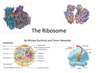
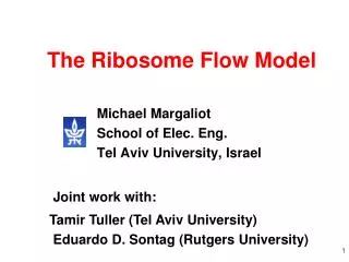
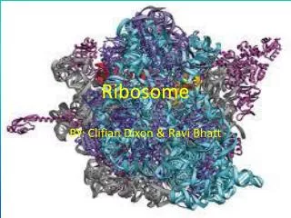
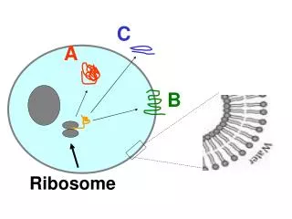
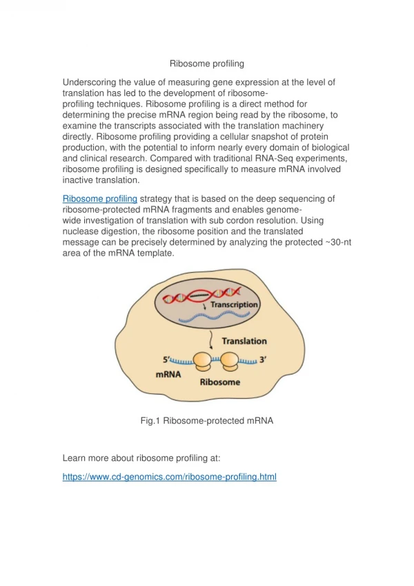
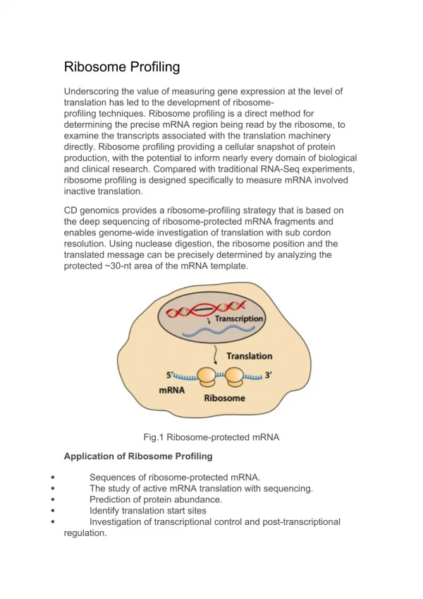
Ribosome
C. A. B. Ribosome. Copy the figure. What is the difference between proteins A, B, C? 2 ) Where in the cell are these proteins synthesized? 3) The preparation of proteins B and C needs more steps than protein A. Why?. Functions of Membrane Proteins:. Transporter. Enzyme. Receptor.


Ribosome
E N D
Presentation Transcript
C A B Ribosome
Copy the figure. • What is the difference between proteins A, B, C? • 2) Where in the cell are these proteins synthesized? • 3) The preparation of proteins B and C needs more steps than protein A. Why?
Functions of Membrane Proteins: Transporter Enzyme Receptor Cell Identity
Functions of Membrane Proteins (B) Transport Proteins Receptors Recognition “Tags” Enzymes
C: A protein that is secreted by exocytosis. For example, Insulin (a hormone)
A Few General Facts: • Humans make about 30,000 different proteins (amino-acid chains). • About 30% of these proteins are connected to the membrane. • About 10% are secreted outside cells. • The rest (60%) are water-soluble and function in the cytoplasm.
C A B Ribosome So, how do proteins get delivered to the membrane?!
Vesicular Transport Stolaf • Watch animation, use handout to mark the steps. • What is the purpose of the process? • What is happening in each step (6 steps)? Vesicular Transport animation
“How Cells Operate?” Work on the handout – coloring, short questions. Can you now tell what each part of the cell is responsible for?
Smooth ER (no ribosomes) Rough ER (with ribosomes)
While you are watching: • What are the roles of: • Cytoskeleton • Nucleus • Ribosomes • Motor proteins Cell Motion Music Click on “Watch video, high” Cell Motion Narrative
Red Blood Cells T-Helper Lymphocyte Signal from another body cell Lymphocyte Lipid Raft: Cell, Stop!
Signal Transduction into cell interior Decent into interior along microfilaments Microfilaments are changing cell shape
Actin filament snapped into two by some enzyme. Microtubules assembly Micro-tubules disassembly - Net “movement”
Kinesin carries vesicle on microtubulue. Contains proteins destined to the outside Microtubule organizing center
Focus shifts from MOTC to the nucleus mRNA shoots out of the nucleus mRNA triggers the assembly of ribosomes
Ribosome translates a new protein that meantime folds up. Protein is sent to the mitochondria Another protein is translated and spinned into the ER
Vesicles carry new protein to Golgi. Golgi vesicles carry protein to cell membrane New protein is released to extra-cellular space
Lipid raft gathers around new protein Membrane proteins rise up Proteins match and connect to opposite arteriole cell
As a result, arteriole cell makes space for white blood to exit. Lymphocyte squeezes out to fight an infection.
Homework: Use page 79. On the cell coloring handout – Fill in the function of each organelle, and specify if it is related to: Protein synthesis and delivery Information center Energy Storage and waste disposal Physical support