Download

1 / 55
670 likes | 1.44k Views
Management of intraductal papillary mucinous neoplasm of pancreas (IPMN) An Update. KOK SIU YAN AMY United Christian Hospital. IPMN. Introduction Classification Investigation Indication for resection Methods of resection Follow-up Prognosis. Introduction. History: 1982

E N D
Management of intraductal papillary mucinous neoplasm of pancreas (IPMN)An Update KOK SIU YAN AMY United Christian Hospital
IPMN • Introduction • Classification • Investigation • Indication for resection • Methods of resection • Follow-up • Prognosis
Introduction • History: 1982 • Described by Ohashi and his colleagues • Incidence ~2.04 per 100 000 • Autopsy studies 25% of cystic pancreatic lesions • 30-50% may become invasive • Accounts for 5-7% of all pancreatic neoplasm Dtsch Arztebl Int. 2011 Nov;108(46):788-94. Intraductal papillary mucinous neoplasia (IPMN) of the pancreas: its diagnosis, treatment, and prognosis. Grützmann R, Post S, Saeger HD, Niedergethmann
Presentation DtschArztebl Int. 2011 Nov;108(46):788-94.. Intraductal papillary mucinousneoplasia (IPMN) of the pancreas: its diagnosis, treatment, and prognosis. Grützmann R, Post S, Saeger HD, Niedergethmann M.
Management guideline First international consensus guideline in 2006 and was revised in 2012
classification • Main duct • Branch duct • Mixed
Investigation • USG • CT • MRI + MRCP • EUS +/- FNAC • ERCP • Intraductal USG or pancreatoscopy
investigation • USG • Size and extent • CT • Diffusely distended pancreatic duct with mucinous filling defects and cystic space occupying lesions • Sensitivity 42%
investigation • MRI with MRCP • Entire pancreatic parenchyma and the intra-abdominal organs • More sensitive to detect mural nodules • Sensitivity 88% Br J Surg. 2009 Jan;96(1):5-20. Preoperative tissue diagnosis for tumours of the pancreas. Hartwig W, Schneider L, Diener MK, Bergmann F, Büchler MW, Werner J.
Investigation • EUS-FNA • CEA • Amylase • Cytology • Molecular analysis • Investigational • Small BD-IPMN
INDICATIONS FOR RESECTION • MD-IPMN • BD-IPMN
2006 guideline: MD-IPMN • Main duct dilatation≥1cm • Frequency of malignancy: 60-92% • No clinical parameters accurately discriminate malignant and non-malignant lesion RESECT ALL MAIN DUCT AND MIXED VARIANT IPMN IF SURGICALLY FIT
2012 guideline: BD-IPMN • High-risk stigmata • Obstructive jaundice in patient with cystic lesion of the head of pancreas • Enhancing solid component within cyst • Main pancreatic duct ≥10mm in size CONSIDER SURGERY IF CLINICALLY APPROPRIATE
2012 guideline: BD-IPMN • Worrisome features • Clinical: pancreatitis • Imaging: • cyst≥3cm • Thickened/enhancing cyst walls • Main duct size 5-9mm • Non-enhancing mural nodule • Abrupt change in calibre of pancreatic duct with distal pancreatic atrophy PERFORM ENDOSCOPIC ULTRASOUND
2012 guideline: BD-IPMN • EUS features: • Definite mural nodule • Main duct features suspicious for involvement • Cytology: suspicious or positive for malignancy CONSIDER SURGERY IF CLINICALLY APPROPRIATE
2012 guideline: MD-IPMN • MD-IPMN • Main duct dilatation≥5mm • 5-9mm worrisom features • Evaluation • No immediate resection
Method of pancreatectomy • According to site and extend of disease • Pancreatoduodenectomy • Distal pancreatectomy • Total pancreatectomy • Limited resection • Excision • Enucleation • Uncinatectomy • Laparoscopy +/- lymph node dissection
follow-up • Non-resected IPMN • Surgically resected IPMN
2006: resected IPMN • Benign: • Yearly CT/MRI • Malignant • 6 monthly CT/MRI • CEA & CA19.9 no value
2012: Resected IPMN • Surgical margin status • normal pancreatic tissue • non-dysplastic changes • low grade dysplasia • moderate grade dysplasia • invasive carcinoma Repeat exam 2-5 years Hx/PE/MRCP half-yearly Identical to PDAC
prognosis • Complete resection of noninvasive IPMN • 5 year survival rate 95% • Complete resection of early stage tumors(T1N0) • 5 year survival rate 60% • Advanced or nodal positive tumors • 5 year survival rate 36%
investigation • High-risk stigmata surgery • Smaller cyst + worrisome features EUS • Cyst >3cm + NO worrisome features EUS if elderly • Cyst ≤3cm + NO worrisome features surveillance
Md-ipmn • High incidence of malignant/invasive lesions (61.6%/43.1%) • Segmental ectatic type/Diffuse type with focal lesions • Diffuse type without focal lesions • Frozen section • Intraductal USG/pancreatoscopy
Bd-ipmn • Mean frequency of malignancy/invasive cancer (25.5/17.7%) • Elderly • Annual malignancy risk 2-3% • High risk factors: • Mural nodules • High grade dysplasia/Positive cytology • Rapidly increasing size • Individual decision
Family history • One 1st degree relative with PDAC 2.3-fold increased risk • high-quality MRI/MRCP or CT and EUS • Malignant stigmata/worrisome features • resection • No malignant stigmata/worrisome features • MRI/MRCP or CT at 3-month intervals • EUS annually for first 2 years
Distinction of BD-IPMN from mcn & other pancreatic cyst • Combination of clinical & imaging characteristics can provide preoperative diagnosis of cyst type • Multidetector CT (MDCT) & MRCP are useful for defining morphology, location, multiplicity, and communication with the MPD • Distinguishing features: multiplicity & visualisation of a connection to MPD • EUS: delineate malignant characteristics eg mural nodules & invasion but operator dependent • Cyst fluid x CEA, amylase & cytology cannot distinguish MCN & IPMN • Molecular analysis for GNAS mutations can distinguish MCN from BD-IPMN
Distinction of BD-IPMN from serous cystic neoplasm (SCN) • 3 morphological patterns: • Polycystic • Honeycomb • Oligocystic Distinguished from SCN with a polycystic or honeycomb pattern by CT or MRCP Differentiation between a small oligocystic SCN & a BD-IPMN may require EUS-FNA with cyst fluid CEA determination
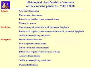
Pancreatic cyst • Neoplastic cysts: • Non-mucinous • Serous cystic neoplasm (SCN) • Solid pseudopapillary neoplasm (SPN) • Mucinous • Mucinous cystic neoplasm (MCN) • Intraductal papillary mucinous neoplasm (IPMN)
MCN • Low prevalence of invasive carcinoma (<15%) • Resection is recommended • Young • Risk of progression • Locations in body and tail • High cost of long-term FU • Distal pancreatectomy • Parenchymal-sparing resection (middle pancreatectomy) • Laparoscopic
EUS-FNA • Apart from imaging, elevated cyst fluid CEA is a marker that distinguishes mucinous from non mucinous cysts, but NOT benign from malignant cysts • A cut off of >/=192-200ng/ml is ~80% accurate for diagnosis of mucinous cyst • Cyst fluid amylase is shown to be not uniformly elevated in IPMN • Fluid cytology may add value especially for evaluation of a small BD-IPMN without “worrisome features”. • High grade epithelial atypia recognised in cyst fluid predicted malignancy in a mucinous cyst with 72% sensitivity in one study and detected 30% more cancers in small IPMN without worrisome features in another study • Some studies showed molecular analysis of cyst fluid may be helpful in distinguishing significant mucinous cysts from indolent cysts that can be conservatively managed • However, in view of the inconclusive evidence, this guideline suggests cyst fluid analysis is still investigational, but is recommended for evaluation of small BD-IPMN without worrisome features only in centres with expertise in EUS-FNA and cytological interpretation
prognosis • Synchronous/metachronous malignant diseases in extra-pancreatic organs • 20-30% • Frequency and location of extra-pancreatic malignancies differs • GI cancer is common in Asia • Skin/breast/prostatic cancers common in US
conclusion • This comprehensive guideline has lowered the criterion for characterising MD-IPMN to MPD dilatation of >5mm without losing specificity for radiologic diagnosis • -high risk stigmata and worrisome features have been defined to stratify risk of malignancy in BD-IPMN and consider resection or increased freq of surveillance • -resection is recommended for all surgically fit patients with MD-IPMN or MCN • Indications for resection of BD-IPMN are more conservative • BD IPMN >3cm without high risk stigmata can be observed without immediate resection
A previous history of diabetes, especially with insulin use, CP, and family history of PDAC are all relevant risk factors for the development of IPMN. • Am J Gastroenterol. 2013 Jun;108(6):1003-9. doi: 10.1038/ajg.2013.42. Epub 2013 Mar 5. • Risk factors for intraductal papillary mucinous neoplasm (IPMN) of the pancreas: a multicentre case-control study.
MR LKF 75/m • Good past health • No history of pancreatitis • No family history of pancreatic cancer • Physical examination: unremarkable
Ultrasound 2.5cm cystic lesion over pancreas What should we do next?
Pancreatic cyst Differential diagnosis • Benign – Pseudocyst – Serous cystic neoplasm (SCN) – Simple cyst, retention cyst, congential, lymphoepithelial cyst • Potentially malignant – Intraductal papillary mucinous neoplasm (IPMN) – Mucinous cystic neoplasm (MCN) – Solid pseudopapillary neoplasm (SPN) – Neuroendocrine tumor • Malignant – Ductal adenocarcinoma
Pancreatic cyst Pathological diagnosis (n = 212) Fernandez-del Castillo et al. Arch Surg 2003
introduction • WHO classification of digestive system 2010 • IPMN with low or intermediate grade dysplasia • IPMN with high grade dysplasia • IPMN with invasive cancer