Download
1 / 21
260 likes | 733 Views
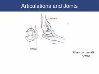
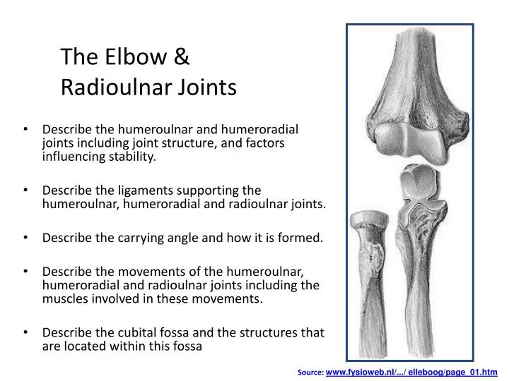
The Elbow & Radioulnar Joints. Describe the humeroulnar and humeroradial joints including joint structure, and factors influencing stability . Describe the ligaments supporting the humeroulnar , humeroradial and radioulnar joints. Describe the carrying angle and how it is formed.

E N D
The Elbow & Radioulnar Joints • Describe the humeroulnar and humeroradial joints including joint structure, and factors influencing stability. • Describe the ligaments supporting the humeroulnar, humeroradial and radioulnar joints. • Describe the carrying angle and how it is formed. • Describe the movements of the humeroulnar, humeroradial and radioulnar joints including the muscles involved in these movements. • Describe the cubitalfossa and the structures that are located within this fossa Source: www.fysioweb.nl/.../ elleboog/page_01.htm
Distal Humerus • Trochlea • Resembles rounded empty spool of thread • Almost complete circle, seperated by thin wall of bone • The groove runs as a spiral around the bone • Articulates with the trochlear notch of the ulna • Capitulum • Hemisphere of bone on anterior inferior surface of distal humerus • Medial border is truncated to form the capitulotrochlear groove • Radial fossa located proximal to capitulum
Distal Humerus Posterior View Anterior View Source: mywebpages.comcast.net/ wnor/lesson4bonesofarm...
Proximal Radius/Ulna • Proximal Ulna • Olecranon process forms the point of the elbow • Coronoid process projects sharply from anterior proximal ulna • Trochlear notch articulates with the trochlea of the humerus. It contains a curved longitudinal ridge that fits into the groove of the trochlea. • Radial notch – depression lateral to inferior edge of trochlear notch • Proximal Radius • Concave superior surface to articulate with capitulum • Cup like depression called the fovea • Distally to the head is the bicipitaltuberosity, it acts as attachment point for biceps
The Carrying Angle • There is a lateral va’L’gus angle between the upper arm and forearm. • This L shaped angle allows the elbow to fit into the depression above the iliac crest (waistline) and aids in carrying heavy loads. • Usually 5° in Men / 10-15° in Women • It is due to the trochlea of the humerus (medial edge) projecting more distally and anteriorly than the lateral edge.
Joint Capsule & Ligaments • A fibrous capsule encloses the elbow joint as well as the superior radioulnar joint. • It is strengthened at the sides by the radial (medial) and ulnar (lateral) collateral ligaments • Its is relatively weak anteriorly and posteriorly
Elbow and R/U Ligaments Adapted from:www.orthogastonia.com/.../ elbow_anatomy.html
Ligaments of the elbow joint Medial collateral Lig • Anterior, posterior and intermediate • Anterior is strongest and associated with common extensor tendon • Triangular Shaped • Protect against valgus stress Lateral Collateral Lig • Less defined than medial ligament • Divided into bundles • one binds with annular ligament • the other attaches to the superior crest of the ulna. • Protects against vargus stress Both are continuous with the joint capsule, are tense in flexion and extension, and strictly limit any abduction, adduction, axial rotation
Ligaments of the Radioulnar Jt • Annular Ligament • Encircles 4/5’s of the radial head • Attached to the anterior and posterior margins of the radial notch. • Flexible – allows the head to rotate i.e. pronate/supinate • Strong – holds the radial head into the radial notch. • Upper fibres strengthened by the radial collateral ligament. • Interoseous Membrane • Strong fibrous sheet which stretches between the the inner surfaces of radius and ulna • Runs from proximal to distal • Helps to absorb forces transmitted from the hands, • Prevent displacement of the radius from the ulna and, • Provides a large surface for muscle attachments. • Also found between the tibia and fibula.
Interosseous Membrane Source: web.sc.itc.keio.ac.jp/.../ A03506001-003.html
Flexion: Biceps Brachii Brachialis Brachioradialis Pronator Teres Extension: Triceps brachii Anconeus Pronation: Pronator Teres Pronator Quadratus Brachioradialis Supination: Supinator Biceps brachii Brachioradialis Musculature
Distal Radioulnar Joint • Head of ulna sits into ulnar notch on radius • Closed inferiorly by triangular articular disc • Stability due to articular disc, interosseous membrane and pronatorquatratus which overlies. • Main movement is supination85° /pronation75 ° • Describe process of supination…
Cubital Fossa Triangular shaped space, bounded laterally by brachioradialis and medially by pronator teres. Main contents include: • Brachial artery • Cephalic and Median cubital veins • Median, radial and lateral cutaneous nerves • Biceps tendon