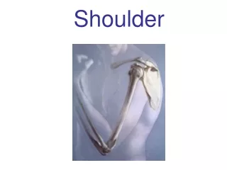
Shoulder Region
Dr. S. Nishan Silva (MBBS). Shoulder Region. Why???. Shoulder Anatomy. Bones Clavicle Scapula Humerus Shoulder vs Shoulder Girdle. Shoulder Anatomy. Joints Sternoclavicular. Shoulder Anatomy. Joints Sternoclavicular Acromioclavicular. Distal Clavicle. Coracoclavicular ligaments

Shoulder Region
E N D
Presentation Transcript
Dr. S. Nishan Silva (MBBS) Shoulder Region
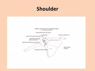
Shoulder Anatomy • Bones • Clavicle • Scapula • Humerus • Shoulder vs Shoulder Girdle
Shoulder Anatomy • Joints • Sternoclavicular
Shoulder Anatomy • Joints • Sternoclavicular • Acromioclavicular
Distal Clavicle • Coracoclavicular ligaments • “Suspensory ligaments of the upper extremity” • Two components: • Trapezoid • Conoid • Stronger than AC ligaments • Provide vertical stability to AC joint
Shoulder Anatomy • Joints • Sternoclavicular • Acromioclavicular • Glenohumeral
Glenohumeral Joint • Most common dislocated joint • Lacks bony stability • Composed of: • Fibrous capsule • Ligaments • Surrounding muscles • Glenoid labrum
Shoulder Anatomy • Ligaments • Acromioclavicular Joint • Acromioclavicular Ligament
Shoulder Anatomy • Ligaments • Glenohumeral Joint • Glenohumeral ligaments • Superior • Middle • Inferior
25% humeral head surface in contact with glenoid Humeral head coverage increased to 75% with glenoid labrum Glenohumeral joint
Shoulder Anatomy • Cartilage • Glenoid labrum
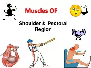
Shoulder Anatomy • Shoulder Girdle Muscles • Trapezius
Major muscles of the trunk Trapezius • Origin: superior nuchal line, external occipital protuberance, ligamentum nuchae and spinous processes of seventh cervical and all thoracic vertebrae • Insertion: lateral third of clavicle, acromion, and spine of scapulartery • Acton: upper fibers elevate scapula, lower fibers depress scapula; if scapula is fixed, one side acting along, draws head toward the same side, and turn face to opposite side; both sides together, draw head directly backward
Major muscles of the trunk Latissimus dorsi • Origin: spinous processes of lower six thoracic and all lumbar vertebrae, median sacral crest, and posterior part of iliac crest. • Insertion: floor of intertubercular groove of humerus. • Action: trunk fixed, extends, adducts and medially rotates arm ; arm fixed, elevates trunk.
Shoulder Anatomy • Shoulder Girdle Muscles • Trapezius • Serratus Anterior
Shoulder Anatomy • Glenohumeral Muscles • Rotator Cuff • Suprispinatus • Infraspinatus • Teres Minor • Subscapularis
Rotator cuff • Subscapularis • Supraspinatus • Infraspinatus • Teres minor
Rotator cuff muscles • Supraspinatus, infraspinatus, teres minor, subscapularis • Form cuff around humeral head • Keep humeral head within joint (counteract deltoid) • Abduction, external rotation, internal rotation
Movement of RC Muscles • Subscapularis is an internal rotator of the arm. • Supraspinatus assists the deltoid in abducting the arm, with its greatest contribution being the initiation of abduction. • Infraspinatus and teres minor muscles both externally rotate the arm.
Teres major • Origin: dorsal surface of inferior angle of scapula • Insertion: crest of lesser tubercle of humerus • Action: medially rotates and adducts arm
Shoulder Anatomy • Glenohumeral Muscles • Latissimus Dorsi • Pectoralis Major
Pectoralis major • Origin: medial half of clavicle,sternum,1th-6th costal cartilages. • Insertion: crest of greater tubercle of humerus. • Action: flexes, adducts and rotates arm medially; arm fixed, elevates trunk; elevates ribs 1-6,aidding in forced inspiration.
Shoulder Anatomy • Glenohumeral Muscles • Latissimus Dorsi • Pectoralis Major • Deltoid
Major muscles of upper limb Deltoid • Origin: lateral third of clavicle, acromion, and spine of scapula • Insertion: deltoid tuberosity of humerus • Action: abducts,flexes and medically rotates, extends, and laterally rotates arm
Shoulder Anatomy • Glenohumeral Muscles • Latissimus Dorsi • Pectoralis Major • Deltoid • Biceps
Biceps strength testing Arms outstretched with palms up at level of shoulder Forced supination of hand with elbow flexed at 90 degrees
Shoulder Anatomy • Glenohumeral Muscles • Latissimus Dorsi • Pectoralis Major • Deltoid • Biceps • Triceps
Shoulder Anatomy • Other structures • Brachial Plexus • Brachial Artery
Brachial plexus Formation: • Five roots: formed by anterior rami of C5-C8 and T1 spinal nerves, roots C5~C7give rise to long thoracic n. • Three trunks • The upper trunk is formed by the joining of root C4,C5,C6. • The middle trunk is the continuation of root C7. • The lower trunk is formed by the joining of root C8 and T1. • Six divisions: above clavicle, trunks form anterior and posterior divisions • Three cords: below clavicle, divisions form three cords that surround the second portion of axillary a.
Position: passes through the scalene fissure to posterosuperior of subclavian artery, then enters the axilla to form lateral, medial and posterior cords Main branches • Lateral cord • Musculocutaneous n. • Lateral root to median n. • Medial cord • Medial root to median n. • Ulnar n. • Medial brachial cutaneous n. • Medial antebrachial cutaneous n.
Posterior cord • radial n. • axillary n. • thoracodorsal n.
Axillary artery • Continuation of subclavian artery at lateral border of first rib • Becomes brachial artery at lower border of teres major • Divided into three parts by overlying pectoralis minor • First portion, above muscle-gives rise to thoracoacromial a. • Second portion, behind muscle-gives rise to lateral thoracic a. • Third portion, below muscle-gives rise to subscapular a. anterior and posterior humeral circumflex a.; the former then divides into throcodorsal a.and circumflex scapular a.
Axillary a. Thoracoacromial a. Lateral pectoral n. Musculocutaneous n. Medial antebrachial cutaneous n. Median n. Ulnar n. Medial brachial cutaneous n. Intercostobrachial n. Thoracodorsal n. & a. Long thoracic n. & lateral thoracic a.
Name the muscles for Horizontal Adduction Pect Major (both) Corachobrachialis Deltoid (anterior) Name the muscles for Horizontal Abduction Deltoid (post) Infraspinatus Teres minor Lats Practice
List the muscles that do flexion of the shoulder Coracobrachialis Pectoralis major (upper to 60°) Anterior Deltoid Practice • List the muscles that do extension of the shoulder • Latissimus dorsi • Teres major • Posterior deltoid • Pectoralis major (lower fibers to neutral)
List the muscles that do adduction of the shoulder Pectoralis major (lower and upper below 90°) Coracobrachialis Latissimus dorsi Teres major Practice • List the muscles that do abduction of the shoulder • Deltoid (all sections) • Supraspinatus • Pectoralis major (upper past 90°)
List the muscles that do internal rotation of the shoulder Subscapularis Latissimus dorsi Teres major Anterior deltoid Pect. major Practice • List the muscles that do external rotation of the shoulder • Infraspinatus • Teres minor • Posterior deltoid
Name the muscle. Coracobrachialis Name the action Adduction of the shoulder Also, flexion and hor. add.