VESSELS general overview
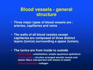
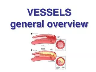
VESSELS general overview. GENERAL STRUCTURE OF VASCULAR WALL. tunica intima subedothe lial layer of connective tissue membrana elastica interna tunica media membrana elastica externa tunica adventitia = tunica externa. General structure of vascular wall. tunica intima tunica media

VESSELS general overview
E N D
Presentation Transcript
GENERAL STRUCTURE OF VASCULAR WALL • tunica intima • subedothelial layer of connective tissue • membrana elastica interna • tunica media • membrana elastica externa • tunica adventitia= tunica externa
General structure of vascular wall • tunica intima • tunica media • tunica adventitia (externa)
Tunica intima • endothelial cells (endotheliocyti) • simple flat/squamous epithelium • on basal lamina • subendothelial layer (stratum subendotheliale) • loose connective tissue • some smooth muscle cells • lamina elastica interna • elastin
Endothelial cells (Endotheliocyti) • mesenchymal origin • zonulae occludentes, desmosomes, nexuses • intermediate filaments, microfilaments (contraction) • corpora multitubularia (Weibel-Palade‘s bodies) • f. VIII – vWF, P-selectin • receptors: adrenergic, histaminic, ADH • synthesis of vasoactive substances: NO, PG
Tunica media • smooth muscle cells • spiral arrangement • elastic a collagen fibers (type III) • lamina elastica externa • only in thicker arteries
Tunica adventitia (externa) • fibroblasts • collagen fibers (type I) • elastic fibers • vasa vasorum • nervi vasorum
Vessel types • arteries (arteriae) - aer + térein • muscular x elastic x mixed • microcirculation: small arteries – less than 1 mm • arterioles (arteriolae) • less than 100 μm • several layers of smooth muscle cells • principal source of peripheral resistance !!! • metarterioly • one smooth muscle cell layer, precapillary sphincter • capillaries (vasa capillaria) • no nerve fibers • endotheliocyte + pericyte(Rouget‘ s cell) • caliber± 7 μm
Vessel types • veins (venae) • few muscle cells, more valvules • venules (venulae) • capacity part of circulation (70% of blood) • lymph vessels(vasa lymphatica) • lymph capillaries (vasa lymphocapillaria) • originate as cul-de-sac • lymphatic trunks and ducts (trunci et ductus lymphatici) • collectors in limbs • valvules
Elastic arteriesaorta, truncus pulmonalis, a. subclavia, axillaris, iliaca, femoralis, thoracica int.
Elastic artery(Arteria elastotypica) • Tunica intima • lamina elastica interna –incoherent • Tunica media • elastic membranes with fenestrations – elastin • smooth muscle cells • lamina elastica externa • Tunica adventitia (externa) • frequent vasa vasorum • supply outer 2/3 of wall
Muscular artery (arteria musculotypica) • Tunica intima • thin • lamina elastica interna – obvious • Tunica media • circular smooth muscle cell layer (up to 40 layerss) • each cell is covered with basal lamina – communication • synthesis of extracellular matrix • lamina elastica externa – several elastic membranes • Tunica externa • nerve bundles – contraction
Arterioles = Arteriolae caliber < 0.5 mm • Tunica intima • Weibel-Palade‘ s bodies within endothelail cells (not in capillaries!) • lamina elastica int. – absent in smallest arterioles • Tunica media • 1-3 layers of smooth muscle cells • lamina elastica ext. – absent • Tunica adventitia (externa) – very thin principal source of peripheral resistance
Sensorystructures in arteries • Glomera supracardiaca (aortica) – sup., medium, inf. - baroreceptors • Sinus caroticus - baroreceptor • thicker, richly innervated tunica adventitia • thinner tunica media • Glomus caroticum - chemoreceptors • oval structures - 3-5 mm • glomus cells – large nucleus, vesicles with catecholamines • shield cells – cover neural endings as glia
Capillaries (Vasa capillaria) • microvascular part of circulation • vas capillare arteriale, intemredium, venosum • site of gas and nutrients exchange • capillary • caliber 7-9 μm • length 1 mm (50 mm in renal glomerulus) • total length approximately 96 000 km • formed by endothelial cells on basal lamina
Capillaries – wall structures • Endothelial cell (Endothelicytus) • Basal lamina (Lamina basalis) • Pericyte (Pericytus; Rouget‘s cells) • mesenchymal cells with long processes • stem, supporting and transporting cell • proper lamina basalis • contractile proteins (replaces tunica media)
Capillary types • somatic capillaries • muscle, conncetive tissue, exocrinne glands (pinocytar vesicle in the wall), nervous tissue (no vesicles) • fenestrated (visceral) capillaries with diaphragms • fenestrations 60-80 nm (quick metabolic exchange) • kidneys, gut, endocrinne glands • fenestrated capillaries without diaphragms • glomeruli in kidney • sinusoids • caliber 30-40 μm, often without lamina basalis • hematopoetic organs – liver, spleen, bone marrow, dental pulp • glomus, glomi n. (vessel glomerule) – ball of fingers, nailbeds, auricle, penis / clitoris, uterus
Capillaries -function • permeability • exchanger vessels (diffusion, pores, fenestration, vesicles) • metabolic function • activation of angiotensin I angiotensin II (lungs) • inactivation of bradykinin, serotonin, prostaglandins • lipolysis • antithrombotic function • inhibition of tissue thromboplastin
Other structures of vascular wall • vasa vasorum • vasa nervorum • nervi vasorum
Other peculiar vascular structures • vas anastomoticum (anastomosis) • vas collaterale (collateral) • rete mirabile = portal system • 2 capillary beds series-connected • anastomosis arteriovenosa (arteriolovenularis) • endothel bulges of intimal cushions with myoepitheloid cells • simple (skin, lungs, kidneys) • composed (glomus coccygeum)
Vessel network arrangement • terminal (retina, spleen, kidney) • functionally terminal (heart, brain) • anastomotic angiogenesis – hypoxia is the strongest factor !
Clinical relevance • aneurysma • atherosclerosis (athere + skleros) • necrosis, infarctus • air embolism in large cervical veins • varices
Blood distribution in organs • heart (coronary arteries) 5% • brain 15% • muscles 15% • viscera 35% • kidneys 20% • skin, skeleton 10% according to Stingl
Developmental arteries • Saccus aorticus (aortal sac) • Aa. arcuum pharyngeorum (pharyngeal arch arteries; „aortal arches“) • 5 pairs develop and change successively • Aorta dorsalis (original 2 merge into 1) • a.a segmentales ventrales ( a. omphalomesenterica, unpaired branches from AA) • truncus umbilicalis ( a. iliaca communis + int.) • a. umbilicalis • aa. segmentales laterales ( paired branches from AA) • aa. intersegmentales dorsolaterales ( branches from a. subclavia) • a. sacralis mediana
Pharyngeal arch arteries derivates • 1st pair – arteria maxillaris + carotis externa • 2nd pair – arteria stapedia • 3rd pair – central – arteria carotis communis – peripheral – arteria carotis interna
Pharyngeal arch arteries derivates • 4th pair • left – part of the arcus aortae • right – a. subclavia dx. • peripheral part of a. subclavia dx.is derived from aorta dorsalis dextra • a. subclavia sin. is NOT derived from the 4th aortic arch but from 7th intersegmental artery
Pharyngeal arch arteries derivates • 5th – Ø • 6th pair • left central left pulmonary artery peripheral ductus arteriosus (Botali) • right central right pulmonary artery peripheral Ø
Arteriae omphalomesentericae(vitellinae) • number of paired arteries • supply yolk sac • develop in vascular supply of gut → truncus coeliacus, arteria mesenterica superior et inferior
Arteriae umbilicales • paired branches • central: truncus umbilicalis from aorta dorsalis • peripheral: within mass of diverticulum allantoicum • to placenta (originally to allantois) in embryonic (connective) stalk or later in umbilical cord • persist as arteriae iliacae internae and vesicales superiores • central: pars patens) • peripheral: ligamentum umbilicale mediale = pars occlusa
Malformation of arteries • Ductus arteriosus patens • Coarctatio aortae • Arcus aortae duplex • Arcus aortae dexter • Arteria lusoria • abnormal origin of the right subclavian artery – obliteration of right aortic arch – origin of 7th segmental artery