Download
1 / 1
10 likes | 111 Views
Investigating Pithecellobium jiringa's effect on glucose absorption to prevent hyperglycemia by analyzing seed and pericarp extracts through bioactivity screening, extraction, and TLC analysis. Results show potential glucose-lowering properties.

E N D
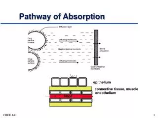
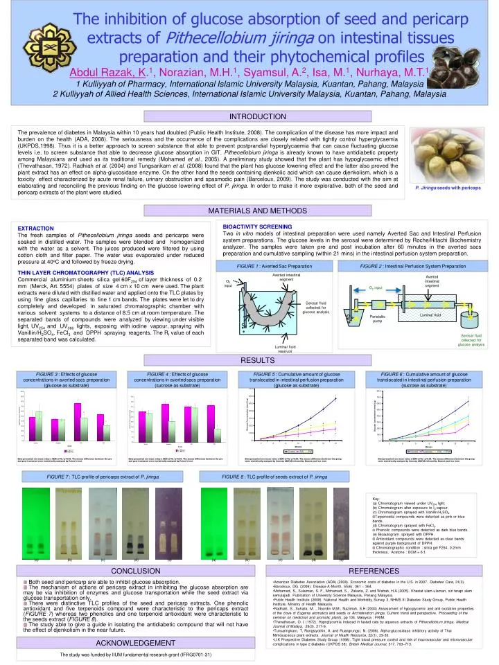
The inhibition of glucose absorption of seed and pericarp extracts of Pithecellobiumjiringa on intestinal tissues preparation and their phytochemical profiles Abdul Razak, K.1, Norazian, M.H.1, Syamsul, A.2, Isa, M.1, Nurhaya, M.T.11 Kulliyyah of Pharmacy, International Islamic University Malaysia, Kuantan, Pahang, Malaysia 2 Kulliyyah of Allied Health Sciences, International Islamic University Malaysia, Kuantan, Pahang, Malaysia Averted intestinal segment O2 input INTRODUCTION The prevalence of diabetes in Malaysia within 10 years had doubled (Public Health Institute, 2008). The complication of the disease has more impact and burden on the health (ADA, 2008). The seriousness and the occurrence of the complications are closely related with tightly control hyperglycaemia (UKPDS,1998). Thus it is a better approach to screen substance that able to prevent postprandial hyperglycaemia that can cause fluctuating glucose levels i.e. to screen substance that able to decrease glucose absorption in GIT. Pithecellobium jiringa is already known to have antidiabetic property among Malaysians and used as its traditional remedy (Mohamed et al., 2005). A preliminary study showed that the plant has hypoglycaemic effect (Thevathasan, 1972). Radhiah et al. (2004) and Tungsarikarn et al. (2008) found that the plant has glucose lowering effect and the latter also proved the plant extract has an effect on alpha-glucosidase enzyme. On the other hand the seeds containing djenkolic acid which can cause djenkolism, which is a toxicity effect characterized by acute renal failure, urinary obstruction and spasmodic pain (Barceloux, 2009). The study was conducted with the aim at elaborating and reconciling the previous finding on the glucose lowering effect of P. jiringa. In order to make it more explorative, both of the seed and pericarp extracts of the plant were studied. Averted intestinal segment O2 input P. Jiringaseeds with pericaps MATERIALS AND METHODS BIOACTIVITY SCREENING Two in vitro models of intestinal preparation were used namely Averted Sac and Intestinal Perfusion system preparations. The glucose levels in the serosal were determined by Roche/Hitachi Biochemistry analyzer. The samples were taken pre and post incubation after 60 minutes in the averted sacs preparation and cumulative sampling (within 21 mins) in the intestinal perfusion system preparation. EXTRACTION The fresh samples of Pithecellobium jiringa seeds and pericarps were soaked in distilled water. The samples were blended and homogenized with the water as a solvent. The juices produced were filtered by using cotton cloth and filter paper. The water was evaporated under reduced pressure at 40oC and followed by freeze drying. THIN LAYER CHROMATOGRAPHY (TLC) ANALYSIS Commercial aluminium sheets silica gel 60F254 of layer thickness of 0.2 mm (Merck, Art. 5554) plates of size 4 cm x 10 cm were used. The plant extracts were diluted with distilled water and applied onto the TLC plates by using fine glass capillaries to fine 1 cm bands. The plates were let to dry completely and developed in saturated chromatographic chamber with various solvent systems to a distance of 8.5 cm at room temperature. The separated bands of compounds were analyzed by viewing under visible light, UV254 and UV366 lights, exposing with iodine vapour, spraying with Vanillin/H2SO4, FeCl3 and DPPH spraying reagents. The Rf value of each separated band was calculated. Serosal fluid collected for glucose analysis FIGURE 1 : Averted Sac Preparation FIGURE 2 : Intestinal Perfusion System Preparation Luminal fluid Peristaltic pump RESULTS FIGURE 3 : Effects of glucose concentrations in averted sacs preparation (glucose as substrate) FIGURE 4 : Effects of glucose concentrations in averted sacs preparation (sucrose as substrate) FIGURE 5 : Cumulative amount of glucose translocated in intestinal perfusion preparation (glucose as substrate) FIGURE 6 : Cumulative amount of glucose translocated in intestinal perfusion preparation (sucrose as substrate) Serosal fluid collected for glucose analysis Luminal fluid reservoir Data presented are mean value ± SEM (n=6).*p<0.05. The means difference between the pre and post-treatment were statistically analyzed by Paired t-test. Data presented are mean value ± SEM (n=6).*p<0.05. The means difference between the pre and post-treatment were statistically analyzed by Paired t-test. Data presented are mean value ± SEM (n=6).*p<0.05. The means difference between the group were statistically analyzed by Oneway ANOVA followed by Dunnet post hoc test. Data presented are mean value ± SEM (n=6).*p<0.05. The means difference between the group were statistically analyzed by Oneway ANOVA followed by Dunnet post hoc test. FIGURE 7 : TLC profile of pericarps extract of P. jiringa FIGURE 8 : TLC profile of seeds extract of P. jiringa • Key: • (a) Chromatogram viewed under UV254 light. • (b) Chromatogram after exposure to I2 vapour. • (c) Chromatogram sprayed with Vanillin/H2SO4. • Terpenoidal compounds were detected as pink or blue bands. • (d) Chromatogram sprayed with FeCl3. • Phenolic compounds were detected as dark blue bands. • (e) Bioautogram sprayed with DPPH. • Antioxidant compounds were detected as clear bands against purple background of DPPH. • Chromatographic condition : silica gel F254, 0.2mm thickness, Acetone : DCM = 6:1. (a) (b) (c) (d) (e) (a) (b) (c) (d) (e) CONCLUSION REFERENCES • Both seed and pericarp are able to inhibit glucose absorption. • The mechanism of actions of pericarp extract in inhibiting the glucose absorption are may be via inhibition of enzymes and glucose transportation while the seed extract via glucose transportation only. • There were distinctive TLC profiles of the seed and pericarp extracts. One phenolic antioxidant and five terpenoids compound were characteristic to the pericaps extract (FIGURE 7) whereas two phenolics and one terpenoid antioxidant were characteristic to the seeds extract (FIGURE 8). • The study able to give a guide in isolating the antidiabetic compound that will not have the effect of djenkolism in the near future. • American Diabetes Association (ADA) (2008). Economic costs of diabetes in the U.S. in 2007. Diabetes Care, 31(3). • Barceloux, DG. (2009). Disease-A-Month. 55(6), 361 – 364. • Mohamed, S., Sulaiman, S. F., Mohamad, S., Zakaria, Z. and Wahab, H.A (2005). Khasiat ulam-ulaman, siri terapi alam semulajadi. Publication of University Science Malaysia, Penang Malaysia. • Public Health Institute (2008). National Health and Morbidity Survey 3, NHMS III Diabetes Study Group, Public Health Institute, Ministry of Health Malaysia. • Radhiah, S., Suhaila, M. ., Noordin M.M., Nazimah, S.H (2004) Assessment of hypoglycemic and anti-oxidative properties of the clove of Eugenia aromatica and seeds or Archidendron jiringa. Current trend and perspective. Proceeding of the seminar on medicinal and aromatic plants, pp 104. Malaysia : FRIM. • Thevathesan, O. I. (1972). Hypoglycemia induced in fasted cats by aqueous extracts of Pithecellobium jiringa. Medical Journal of Malaya, 26(3), 217-9. • Tunsaringkarn, T, Rungsiyothin, A. and Ruangrungsi, N. (2008). Alpha-glucosidase inhibitory activity of Thai Mimosaceous plant extracts. Journal of Health Resource, 22(1), 29-33. • U.K Prospective Diabetes Study Group (1998). Tight blood pressure control and risk of macrovascular and microvascular complications in type 2 diabetes (UKPDS 38). British Medical Journal, 317, 703–713. ACKNOWLEDGEMENT The study was funded by IIUM fundamental research grant (IFRG0701-31)