Download
1 / 29
310 likes | 563 Views
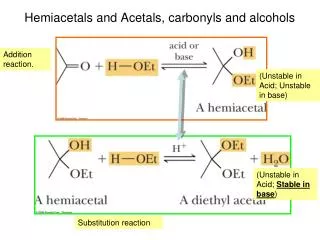
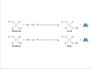
Figure 7.16 Acetals and ketals can be formed from hemiacetals and hemiketals, respectively. Sugar acetals are called glycosides. - glycosides are stable compounds - they do not mutarotate.

E N D
Figure 7.16 Acetals and ketals can be formed from hemiacetals and hemiketals, respectively.
Sugar acetals are called glycosides. - glycosides are stable compounds - they do not mutarotate
Figure 7.18 The structures of several important disaccharides. Note that the notation -HOH means that the configuration can be either or . If the -OH group is above the ring, the configuration is termed . The configuration is if the -OH group is below the ring as shown. Also note that sucrose has no free anomeric carbon atoms.
a nonreducing sugar Fig. 7-18a, p.216
Fehlings reaction for aldehyde(can also detect ketones after tautamerization)
Trehalose – A Natural Protectant for Bugs Insects use an open circulatory system to circulate “hemolymph” (insect blood). The “blood sugar” is trehalose, an unusual, nonreducing disaccharide. Trehalose may act as a natural cryoprotectant, protecting the insect from damage due to freezing temperatures.
Lactose tolerance is widespread among people who come from areas that have a long history of dairy farming - including Northern Europe, the Middle East and India - or who rely extensively on milk in their diets, such as the Fulani in West Africa or the Masai in East Africa. But adults from other areas - such as East Asia and much of Africa - remain lactose intolerant, and find it difficult to digest milk.
A Variety of Higher Oligosaccharides Occur in Nature Oligosaccharides occur widely as components of antibiotics (derived from various sources). Figure 7.19 Erythromycin is an antibiotic produced by a strain of Streptomyces erythreus.
A Variety of Higher Oligosaccharides Occur in Nature Oligosaccharides occur widely as components of antibiotics (derived from various sources). Figure 7.19 Streptomycin is an oligosaccharide produced by Stretomyces griseus.
7.4 What is the Structure and Chemistry of Polysaccharides? Functions: storage, structure, recognition Nomenclature for polysaccharides is based on their composition and structure Homopolysaccharide – a polysaccharide that contains only one kind of monosaccharide Heteropolysaccharide – a polysaccharide made of several monosaccharides Starch and glycogen are storage molecules Chitin and cellulose are structural molecules Cell surface polysaccharides are recognition molecules
Starch A plant storage polysaccharide Two forms: amylose and amylopectin Most starch is 10-30% amylose and 70-90% amylopectin Branches in amylopectin every 12-30 residues Amylose has alpha(1,4) links, one reducing end The branches in amylopectin are α(1→6).
Amylose and Amylopectin are energy storage molecules in plants Figure 7.20 Amylose and amylopectin are two forms of starch. Amylopectin is highly branched, with branches occurring every 12 to 30 residues.
The Structure of Amylose Amylose is poorly soluble in water, but forms micellar suspensions In these suspensions, amylose is helical Iodine fits into the helices to produce a blue color
The Structure of Amylose Figure 7.21 Suspensions of amylose in water adopt a helical conformation. Iodine (I2) can fit into the middle of the amylose helix to give a blue color that is characteristic and diagnostic for starch.
The Phosphorylase Reaction Releases Glucose Units for Metabolic Energy Figure 7.22 The starch phosphorylase reactions cleaves glucose residues from amylose, producing α-D-glucose-1-phosphate, an energy source for the organism.
Glycogen The glucose storage device in animals Glycogen constitutes up to 10% of liver mass and 1-2% of muscle mass Glycogen is stored energy for the organism Only difference from amylopectin: the frequency of branching Alpha(1,6) branches every 8-12 residues Like amylopectin, glycogen gives a red-violet color with iodine
Dextrans A small but significant difference from starch and glycogen If you change the main linkages between glucose from alpha(1,4) to alpha(1,6), you get a new family of polysaccharides - dextrans Branches can be (1,2), (1,3), or (1,4)
Roles for Dextrans Dextrans formed by bacteria are components of dental plaque Dextrans in plaque presumably provide protection for oral bacteria Cross-linked dextrans are used as "Sephadex" gels in column chromatography These gels, used to separate biomolecules on the basis of size, are up to 98% water!
Structural Polysaccharides The composition of structural polysaccharides is similar to storage polysaccharides But small structural differences greatly influence properties Starch and glycogen linkages consist primarily of α(1→4) linkages. Cellulose consists of β(1→4) linkages
Cellulose Provides Physical Structure and Strength to Plants Cellulose is a structural polysaccharide It is the most abundant natural polymer in the world It is found in the cell walls of nearly all plants The wood and bark of trees are insoluble, highly organized structures formed from cellulose and lignin Cotton is almost pure cellulose Cotton acetates, made from the action of acetic anhydride on cellulose, are used in dresses, lingerie, and other clothing
The Structural Difference Between Amylose and Cellulose Figure 7.23 (a) Amylose prefers a helical conformation (due to its bent α(1→4) linkages. (b) Cellulose, with β(1→4) linkages, can adopt a fully extended conformation.
The Structure of Cellulose Figure 7.24 The structure of cellulose, showing the hydrogen bonds (blue) between the sheets, which strengthen the structure. Intrachain H-bonds in red; interchain H-bonds in green.
How Do Ruminant Animals Digest Cellulose? Figure 7.25 Giraffes, cattle, deer, and camels are ruminant animals that are able to metabolize cellulose, thanks to bacterial cellulase in the rumen, a large first compartment in the stomach of a ruminant.
Other Structural Polysaccharides Chitin – found in the exoskeletons of crustaceans, insects and spiders, and cell walls of fungi It is similar to cellulose, but C-2s are N-acetyl Cellulose strands are parallel; chitins can be parallel or antiparallel
Structures of cellulose, chitin and mannan Figure 7.26 Like cellulose, chitin and mannan form extended ribbons and pack together efficiently, taking advantage of multiple hydrogen bonds.
Other Structural Polysaccharides Alginates – Ca2+-binding polymers in algae Agarose and agaropectin - galactose polymers Glycosaminoglycans - repeating disaccharides with amino sugars and negative charges
The Structure of Agarose in Solution • Agarose, a component of agar obtained from marine red algae, is a chain of alternating D-galactose and 3,6-anhydro-L-galactose. • Agarose gels are used in laboratories to separate biomolecules on the basis of size Figure 7.27 The favored conformation of agarose in water is a double helix with a threefold screw axis.