Download
1 / 39
390 likes | 570 Views
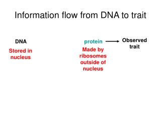
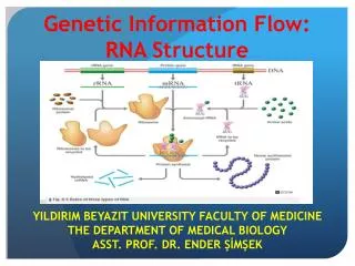
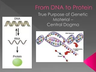
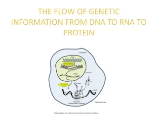
THE FLOW OF GENETIC INFORMATION FROM DNA TO RNA TO PROTEIN. 10.6 The DNA genotype is expressed as proteins, which provide the molecular basis for phenotypic traits The information constituting an organism’s genotype Is carried in its sequence of its DNA bases

E N D
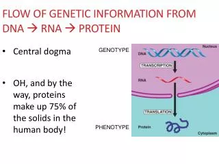
THE FLOW OF GENETIC INFORMATION FROM DNA TO RNA TO PROTEIN • 10.6 The DNA genotype is expressed as proteins, which provide the molecular basis for phenotypic traits • The information constituting an organism’s genotype • Is carried in its sequence of its DNA bases • A particular gene, a linear sequence of many nucleotides • Specifies a polypeptide
DNA Transcription RNA Translation Protein Figure 10.6A • The DNA of the gene is transcribed into RNA • Which is translated into the polypeptide
10.7 Genetic information written in codons is translated into amino acid sequences • The “words” of the DNA “language” • Are triplets of bases called codons • The codons in a gene • Specify the amino acid sequence of a polypeptide
DNA molecule Gene 1 Gene 2 Gene 3 DNA strand A A A C A C G G A A C A Transcription U U U G U G C C U U G U RNA Codon Translation Polypeptide Amino acid Figure 10.7
Second base U C A G U UAU UGU UGC UGA Stop UUU UCU Cys Phe Tyr UUC UAC C UCC Ser U UCA UUA UAA Stop A Leu UCG UAG Stop UGG Trp G U CAU CGU CUU CCU His C CAC CGC CUC CCC C Pro Arg Leu CUA CCA CAA CGA A Gln CAG CGG CUG CCG G Third base First base U ACU AUU AAU AGU Ser Asn ACC AGC AUC AAC Ile C A Thr AUA AGA ACA AAA A Lys Arg Met or start ACC AGG AAG AUG G U GUU GAU GGU GCU Asp C GGC GCC GUC GAC Gly Ala G Val GUA GCA GGA GAA A Glu GUG GCG GGG GAG G Figure 10.8A • 10.8 The genetic code is the Rosetta stone of life • Nearly all organisms • Use exactly the same genetic code UUG
Strand to be transcribed T A C T T C A A A A T C DNA A T G A A G T T T T A G Transcription G U U U A G A U A A G U RNA Startcondon Stopcondon Translation Met Polypeptide Lys Phe Figure 10.8B • An exercise in translating the genetic code
RNA nucleotides RNA polymerase A C C A T T A U T C T G U G A C A U C C A C C A G A T T T A G G Direction of transcription Template Strand of DNA Figure 10.9A Newly made RNA • 10.9 Transcription produces genetic messages in the form of RNA • A close-up view of transcription
In the nucleus, the DNA helix unzips • And RNA nucleotides line up along one strand of the DNA, following the base pairing rules • As the single-stranded messenger RNA (mRNA) peels away from the gene • The DNA strands rejoin
RNA polymerase DNA of gene Promoter DNA Terminator DNA Area shown In Figure 10.9A Growing RNA Completed RNA RNA polymerase Figure 10.9B • Transcription of a gene 1 Initiation 2 Elongation 3 Termination
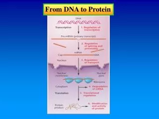
Exon Intron Exon Intron Exon DNA Transcription Addition of cap and tail Cap RNA transcript with cap and tail Introns removed Tail Exons spliced together mRNA Coding sequence Nucleus Cytoplasm Figure 10.10 • 10.10 Eukaryotic RNA is processed before leaving the nucleus • Noncoding segments called introns are spliced out • And a cap and a tail are added to the ends
10.11 Transfer RNA molecules serve as interpreters during translation • Translation • Takes place in the cytoplasm
0 Amino acid attachment site Hydrogen bond RNA polynucleotide chain Anticodon Figure 10.11A • A ribosome attaches to the mRNA • And translates its message into a specific polypeptide aided by transfer RNAs (tRNAs)
Amino acid attachment site Anticodon Figure 10.11B, C • Each tRNA molecule • Is a folded molecule bearing a base triplet called an anticodon on one end • A specific amino acid • Is attached to the other end
tRNAmolecules Growingpolypeptide Largesubunit mRNA Small subunit Figure 10.12A • 10.12 Ribosomes build polypeptides • A ribosome consists of two subunits • Each made up of proteins and a kind of RNA called ribosomal RNA
The subunits of a ribosome • Hold the tRNA and mRNA close together during translation tRNA-binding sites Largesubunit Next amino acid to be added to polypeptide Growing polypeptide tRNA mRNA-binding site mRNA Smallsubunit Codons Figure 10.12B, C
Start of genetic message End Figure 10.13A • 10.13 An initiation codon marks the start of an mRNA message
Large ribosomalsubunit Met Met Initiator tRNA P site A site U C U A C A A U G AUG Startcodon Small ribosomalsubunit mRNA 1 2 Figure 10.13B • mRNA, a specific tRNA, and the ribosome subunits • Assemble during initiation
10.14 Elongation adds amino acids to the polypeptide chain until a stop codon terminates translation • Once initiation is complete • Amino acids are added one by one to the first amino acid
1 Codon recognition 2 Peptide bondformation Translocation 3 • Each addition of an amino acid • Occurs in a three-step elongation process Aminoacid Polypeptide P site A site Anticodon mRNA Codons mRNAmovement Stopcodon New Peptidebond Figure 10.14
The mRNA moves a codon at a time • And a tRNA with a complementary anticodon pairs with each codon, adding its amino acid to the peptide chain
Elongation continues • Until a stop codon reaches the ribosome’s A site, terminating translation
10.15 Review: The flow of genetic information in the cell is DNARNAprotein • The sequence of codons in DNA, via the sequence of codons • Spells out the primary structure of a polypeptide
1 mRNA is transcribed from a DNA template. 2 Each amino acidattaches to its propertRNA with the help of aspecific enzyme and ATP. 3 Initiation ofpolypeptide synthesis The mRNA, the first tRNA, and the ribosomal subunits come together. 4 Elongation A succession of tRNAsadd their amino acids to the polypeptide chain as the mRNA is moved through the ribosome, one codon at a time. 5 Termination The ribosome recognizes a stop codon. The poly-peptide is terminated and released. • Summary of transcription and translation DNA Transcription mRNA RNApolymerase Amino acid Translation Enzyme ATP tRNA Anticodon Largeribosomal subunit InitiatortRNA Start Codon Smallribosomal subunit mRNA New peptidebond forming Growingpolypeptide Codons mRNA Polypeptide Figure 10.15 Stop codon
Normal hemoglobin DNA Mutant hemoglobin DNA C A T T T C mRNA mRNA G A A G U A Normal hemoglobin Sickle-cell hemoglobin Glu Val Figure 10.16A • 10.16 Mutations can change the meaning of genes • Mutations are changes in the DNA base sequence • Caused by errors in DNA replication or recombination, or by mutagens
Normal gene U G C U U C A G A A U G A G G mRNA Met Lys Gly Protein Phe Ala Base substitution A A G A U G C A U G A G U U C Lys Met Phe Ser Ala Missing U Base deletion G G C G A C A U A U G A G U U Figure 10.16B Lys Ala His Met Leu • Substituting, inserting, or deleting nucleotides alters a gene • With varying effects on the organism
MICROBIAL GENETICS • 10.17 Viral DNA may become part of the host chromosome • Viruses • Can be regarded as genes packaged in protein
When phage DNA enters a lytic cycle inside a bacterium • It is replicated, transcribed, and translated • The new viral DNA and protein molecules • Then assemble into new phages, which burst from the host cell
In the lysogenic cycle • Phage DNA inserts into the host chromosome and is passed on to generations of daughter cells • Much later • It may initiate phage production
Phage Attachesto cell Bacterialchromosome Phage DNA Cell lyses,releasing phages Phage injects DNA Many celldivisions Lytic cycle Lysogenic cycle Phages assemble Lysogenic bacterium reproducesnormally, replicating the prophageat each cell division Phage DNAcircularizes Prophage OR Phage DNA inserts into the bacterialchromosome by recombination New phage DNA andproteins are synthesized Figure 10.17 • Phage reproductive cycles 1 7 2 4 3 5 6
Membranousenvelope RNA Protein coat Figure 10.18A Glycoprotein spike CONNECTION • 10.18 Many viruses cause disease in animals • Many viruses cause disease • When they invade animal or plant cells • Many, such as flu viruses • Have RNA, rather than DNA, as their genetic material
1 Entry 2 Uncoating RNA synthesisby viral enzyme 3 RNA synthesis(other strand) 4 Proteinsynthesis 5 6 Assembly • Some animal viruses • Steal a bit of host cell membrane as a protective envelope • Can remain latent in the host’s body for long periods Glycoprotein spike VIRUS Protein coat Viral RNA(genome) Envelope Plasma membraneof host cell Viral RNA(genome) mRNA Template New viralgenome Newviral proteins Exit 7 Figure 10.18B
RNA Protein Figure 10.19 CONNECTION • 10.19 Plant viruses are serious agricultural pests • Most plant viruses • Have RNA genomes • Enter their hosts via wounds in the plant’s outer layers
Colorized TEM 370,000 Colorized TEM 50,000 Figure 10.20A, B CONNECTION • 10.20 Emerging viruses threaten human health
Envelope Glycoprotein Protein coat RNA (two identical strands) Reverse transcriptase Figure 10.21A • 10.21 The AIDS virus makes DNA on an RNA template • HIV, the AIDS virus • Is a retrovirus
Viral RNA CYTOPLASM NUCLEUS Chromosomal DNA RNAstrand Double-strandedDNA Provirus DNA Viral RNAand proteins RNA Figure 10.21B • Inside a cell, HIV uses its RNA as a template for making DNA • To insert into a host chromosome 1 2 3 4 5 6
Mating bridge DNA enters cell Phage Phage Fragment of DNAfrom anotherbacterial cell Fragment of DNA fromanotherbacterial cell(former phagehost) Sex pili Bacterial chromosome (DNA) Recipient cell(“female”) Donor cell(“male”) Figure 10.22A–C • 10.22 Bacteria can transfer DNA in three ways • Bacteria can transfer genes from cell to cell by one of three processes • Transformation, transduction, or conjugation
Degraded DNA Crossovers Donated DNA Recombinantchromosome Recipient cell’schromosome Figure 10.22D • Once new DNA gets into a bacterial cell • Part of it may then integrate into the recipient’s chromosome
10.23 Bacterial plasmids can serve as carriers for gene transfer • Plasmids • Are small circular DNA molecules separate from the bacterial chromosome
F factor (plasmid) F factor (integrated) Male (donor) cell Male (donor) cell Origin of F replication Bacterial chromosome Bacterial chromosome F factor starts replication and transfer F factor starts replication and transfer of chromosome Recipient cell Plasmid completes transferand circularizes Only part of the chromosome transfers Plasmids Colorized TEM 2,000 Recombination can occur Cell now male Figure 10.23A–C • Plasmids can serve as carriers • For the transfer of genes