Download

1 / 45
550 likes | 1.86k Views
Abdominal Pain/Abdominal Mass. Melissa L. Hughes Scott Q. Nguyen, M.D. Celia M. Divino, M.D. Department of Surgery Mount Sinai School of Medicine. HPI Mrs.Masseo. Mrs. Masseo is a 63-year-old female with PMH of HTN, DM, s/p laparotomy for peptic ulcer disease seven years ago

E N D
Abdominal Pain/Abdominal Mass Melissa L. Hughes Scott Q. Nguyen, M.D. Celia M. Divino, M.D. Department of Surgery Mount Sinai School of Medicine
HPI Mrs.Masseo • Mrs. Masseo is a 63-year-old female with PMH of HTN, DM, s/p laparotomy for peptic ulcer disease seven years ago • Presents to ER with one day history of sudden, worsening abdominal pain associated with nausea, two episodes of vomiting, and abdominal distension
What other information would you want regarding this patient’s history?
Other Pertinent HPI Patient had noticed a bulging from her mid abdomen beneath the surgical scar for the past several months. It was not initially painful, became larger when she coughed, and would go away when she was lying down After an acute coughing episode the morning prior to admission, patient reported that she suddenly experienced severe pain in her mid abdomen that was constant and accompanied by an increase in size of the midline bulge which did not go away when she tried to lie down No flatus or bowel movement over the past day, several episodes of vomiting, and subjective fevers
Other Pertinent History • PMH: Poorly controlled HTN and DM for the past 20 years • PSH: Appendectomy at age 35, laparotomy 7 years ago for PUD • Meds: lisinopril, insulin, nexium, aspirin • Allergies: NKDA • Social history: 1.5 packs of cigarettes a day for the past 40 years
Physical Exam Ill-appearing, obese woman in severe pain BP 100/60 HR 115 Temp 38.2 C RR 24 HEENT: oral mucosa dry Heart: tachycardic, regular rhythm Lungs: clear to auscultation bilaterally Abdomen: obese abdomen, healed midline laparotomy and RLQ scars, hypoactive bowel sounds, moderate distension, firm, tender softball size mass at midline scar with erythema of the overlying skin. No rebound or guarding in remaining abdomen Guaiac positive stool
Differential Diagnosis • Incarcerated ventral hernia • Small/large bowel obstruction- secondary to adhesions, volvulus, neoplasm • Abdominal wall tumor • Abdominal wall abscess
Lab results, Mrs. Masseo 10 134 94 40 15 350 190 3.3 20 1.7 30.1 n% 89 LFTs, amylase, lipase, and coags- WNL
Lab Findings Pre-renal azotemia secondary to dehydration Leukocytosis from infection/inflammatory process
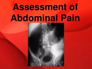
Obstructive Series Describe the X-ray findings
Xray Interpretation • No free air noted on CXR • No significant small bowel dilatation • Air in right colon • No small bowel obstruction
If this patient had bowel obstruction secondary to an incarcerated loop of small bowel in the ventral hernia, then why are there no signs of small bowel obstruction on Xray?Is there another study which may help?
CT Interpretation • Transverse colon incarcerated in ventral abdominal wall hernia • Soft tissue stranding in subcutaneous fat around incarcerated hernia • Absence of enteric contrast past area of incarceration with collapse of left colon consistent with complete large bowel obstruction
Hospital Course Immediate resuscitation with IV fluids, foley catheter, NG tube decompression and pre-op antibiotics Patient taken to the OR for incarcerated hernia with suspected strangulated bowel Exploratory laparotomy performed using previous midline incision Found to have ischemic loop of transverse colon twisted upon itself, herniating through a 4cm abdominal wall defect Segment of ischemic bowel was resected and primary anastomosis performed Hernia repaired primarily, skin was left open
Hospital Course Patient did well post-operatively without complications POD #4: regained bowel function POD #6: tolerated normal diet POD#7: discharged home
What is the problem with repairing this patient’s hernia primarily? Would you want to use mesh in this situation?
Primary repair of Ventral (Incisional) Hernia Recurrence of a ventral hernia is a common problem in primary suture repair, whereas repair with prosthetic mesh often has lower recurrence rates However, in a patient with strangulated, ischemic bowel who undergoes a bowel resection, inserting mesh into a contaminated field increases risk of infection of the mesh and ultimate need for reoperation and removal
Follow-up Patient seen at follow-up appointment 6 months later and was found to have another reducible hernia through the same 4cm abdominal wall defect Patient denied any abdominal pain, distension, nausea, vomiting, or fevers
Discuss treatment options for repair of recurrent incisional hernias • Discuss pre-operative preparation
Follow-up • Patient taken back to the OR for elective ventral hernia repair • Open hernia repair performed using non-absorbable mesh in an under-lay fashion • Patient continues to do well two years after elective repair without any signs or symptoms of recurrence
Incisional Hernia Discussion Hernias that occur at a prior abdominal incision site (includes post laparotomy hernias, parastomal hernias, and trocar site hernias) Incisional hernias reported in up to 20% of patients undergoing laparotomy with modern rates ranging from 2-11% Approximately 100,000 ventral incisional hernia repairs performed each year in U.S. Most present within 12 months post-laparotomy although as many as 1/3 may present 5-10 years later
What are the risk factors for developing an incisional hernia?
Risk Factors • Patient-related factors: advanced age, malnutrition, diabetes mellitus, cigarette smoking, corticosteroids, conditions that increase intra-abdominal pressure like obesity ascites, or chronic cough • Surgery-related factors: wound or intraabdominal infection, closure of abdomen under tension, type and location of incision (vertical midline incision more prone to incisional hernia than transverse), lack of mesh overlap at hernia edges (bridge technique)
Clinical Manifestations and Diagnosis Bulge in abdominal wall at or near surgical scar Discomfort aggravated by coughing or straining Enlarges over time leading to pain, bowel obstruction, incarceration, and strangulation In large hernias, the skin may present with ischemic or pressure necrosis resulting in ulceration Usually easy to identify on exam, with palpable edges of fascial defect In obese patients with suspected incisional hernias the surgeon should have a low threshold for obtaining a CT abdomen as the clinical exam is very unreliable
Treatment Treatment includes two general types of operative repair: primary suture repair and prosthetic mesh repair Recurrence rates for non-prosthetic repair can be as high as 50% or more, whereas mesh repair is associated with significantly lower recurrence rates
Primary Repair Usually performed for hernia defects less than 4 cm in diameter, with strong, viable surrounding tissue using an interrupted layer of nonabsorbable sutures Some studies have suggested that even these small hernias may have a substantially lower recurrence rate after mesh repair Separation of components is a technique that utilizes the body’s own tissues for hernia repair, avoids the use of a foreign body, and in experienced hands may have very good results
Prosthetic Repair For large hernias, or hernias associated with multiple small defects, mesh should be placed by open or laparoscopic approach Mesh provides tension-free repair by avoiding the recreation of tension by fascial apposition. In large hernias with loss of domain , fascial apposition may not even be possible. Much improved recurrence rates over primary repair
Prosthetic Repair • Many different prosthetic materials available today for hernia repair but limited evidence and comparative studies exist • Bioabsorbable meshes have become popular and may be used in an infected field but should not be regarded as permanent hernia repair as high rates of recurrence/ dilatation have recently been described • Many techniques for mesh placement: (ex) Rives-Stoppa repair where mesh is placed in retrorectus space, laparoscopic repair with mesh placement intraabdominally behind the rectus and peritoneum, open in-lay, on-lay and under-lay mesh repairs. • Technique may be paramount in recurrence rates
Complications Recurrence: As high as 30-50% in primary suture repair, 5-35% in open mesh repair, and 0-11% in laparoscopic mesh repair Wound infections are more common after open repair compared to laparoscopic Mesh infection often necessitates removal of mesh but can occasionally be treated with IV antibiotics and local wound care Erosion of mesh into bowel with development of enterocutaneous fistulas Bowel obstruction/ileus
References Feldman LS, et al. Laparoscopic Hernia Repair. ACS Surgery: Principles and Practice. Chapter 5, Section 28. 2003 Fitzgibbons RF, et al. Open Hernia Repair. ACS Surgery: Principles and Practice. Chapter 5, Section 27. 2003 Townsend CM. Sabiston Textbook of Surgery. 17th edition Zinner, MJ, et al. Postoperative Ventral Wall (Incisional) Hernia. Maingot’s Abdominal Operations. Chapter 5. Hernias. 11th edition
Acknowledgment The preceding educational materials were made available through theASSOCIATION FOR SURGICAL EDUCATION In order to improve our educational materials wewelcome your comments/ suggestions at: feedbackPPTM@surgicaleducation.com