ARTERIOVENOUS MALFORMATION
ARTERIOVENOUS MALFORMATION. AVM-Introduction. Vascular malformation: AVM Venous malformation Cavernous malformation Capillary telangiectasia AVF. AVM-introduction. Most dangerous vascular malformation Congenital lesion Abnormal collection of vessels

ARTERIOVENOUS MALFORMATION
E N D
Presentation Transcript
AVM-Introduction Vascular malformation: • AVM • Venous malformation • Cavernous malformation • Capillary telangiectasia • AVF
AVM-introduction • Most dangerous vascular malformation • Congenital lesion • Abnormal collection of vessels wherein arterial blood flows directly into draining veins without the normal capillary beds • Feeding arteries/ Nidus/ Draining veins • Static/ Grow/ Regress
AVM-Presentation • Hemorrhage(50%) • Seizure • Mass effect • Ischemia; steal phenomenon • Headache • Bruit • HCP • Peds: hydrocephalus, heart failure
www.brain-surgery.com AVM-Hemorrhage • Peak age: 15-20 y/o • 10 % mortality; 30-50% morbidity • ICH(80%)/IVH/SAH • Risk of hemorrhage: High feeding a. pressure/V. outflow obstruction/Size/Location/Aneurysm/ Pregnancy
Hemorrhage related to AVM size • Small AVMs are more lethal than larger ones • Small AVMs tends to present more often as hemorrhage than do larger ones 1 • Small AVMs are thought to have much higher pressure in feeding artery 1, 2 1. Crawford P M, West C R, et al: Arteriovenous Malformation: Natural History in Unoperated Patients. J Neurol Neurosuurg Psy 49:1-10,1986 2. Spetzler R F, Hargraves R W, et al: Relationship of Perfusion Pressure and Size to Risk of Hemorrhage from Arteriovenous Malformations. Neurosurgery 37: 851-5, 1995
Annual & Lifetime risk of Hemorrhage • Lifelong risk of bleeding: 2-4% per yr • A study of 166 symptomatic AVMs with 24 year follow-up found the risk of major bleeding was constant at 4% per year, independent of whether the AVM presented with or without hemorrhage 3 • The AVM Study Group: Annual rate of rehemorrhage was 18% among pts who had hemorrhage at presentation; 2% among pts with no history of bleeding (306 cases) 4 • Rebleeding rate significantly lower than aneurysms. 3. Ondra SL, Troupp H, et al: The natural history of symptomatic cerebral arteriovenous malformation: A 24-year follow-up assessment. J Neurosurg 25:387-91, 1990
AVMs & Associated Aneurysms • 7% of pts with AVMs have aneurysms • 75% are located on major feeding artery; probably from increased flow 1 • The symptomatic one is treated first • Although 66% of related aneurysms will regress following AVM removal, this does not always occur 4 4. Cunha M J, Stein B M, et al: The Treatment of Associated Intracranial Aneurysm and Arteriovenous Malformations. J Neurosurg 77: 853-9, 1992.
Hemodynamic Effects of AVM Pre-op effects: • Steal phenomenon • AVM & aneurysm • High-flow angiopathy 7 Post-op effects: • Normal perfusion pressure breakthrough • Occlusive hyperemia 7. Pile Spellman JM, Baker KF, et al: High flow angiopathy: cerebral blood vessel changes in chronic arteriovenous malformation. Am J Neuroradiol 1986; 7:811-5
Cerebral Steal Phenomenon • Autoregulation curve shifts to left • Despite cerebral arterial hypotension, focal neurological deficits are rare(<10%) • More likely to be local mass effect
Normal perfusion pressure breakthrough(NPPB) • Peri-/Post-op swelling or hemorrhage • Loss of autoregulation4 ?5 • Less than 5% • Should be diagnosis of exclusion • Mx: prevent post-op hypertension 4. Spetzler R F, Wilson C B, et al: Normal perfusion breakthrough theory. Clin Neurosurg 25: 651-72, 1978 5. Young W L, Kader A, et al: Pressure autoregulation is intact after arteriovenous malformation resection. Neurosurgery 32: 491-7, 1993
Evaluation-MRI • Flow void on T1WI or T2WI • Feeding arteries • Nidus • Draining veins
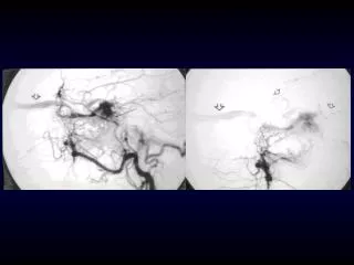
Evaluation-Angiography • Tangle of vessels • Large feeding artery • Large draining veins • Not all AVMs show up on angiography! Angiographically occult vascular malformation (AOVM)
Evaluation-Grading • Spetzler-Martin grade • Outcome based on Spetzler-Martin grade: 100 consecutive cases operated by Spetzler
Treatment • Multidisciplinary approach • Primary goal: decrease the risk of bleeding 1) Surgery: mainstay 2) Stereotactic Radiosurgery (SRS): high-risk for surgery 3) TAE: adjunct to 1) & 2)
Surgery American Stroke Association recommends: • Low grade ( I & II )- surgery alone • Higher grade(>III)-TAE before surgery • Eliminates risk of bleeding immediately, seizure controls improves • Invasive, risk of surgery
Surgery • Pre-op propranolol 20mg po QIDx3d to minimize post-op normal perfusion pressure breakthrough (NPPB) • Peri-op labetalol to keep MAP 70-80mmHg
Surgery • Craniotomy • Dural opening • Identify the borders • Cautery of feeding arteries
Surgery • Deep dissection of the nidus • Securing the ventricle • Obliterate the draining veins • Final removal of AVM • Post-resection BP challenge Hemostasis/ Residual nidus/ Areas prone to NPPB • Immediate post-op/ Peri-op angiography
Intra-Op Complication • Premature division of venous drainage • Extensive bleeding along the deep margin • Post-resection NPPB/ Residual AVM • Pack the wall with Avitene & Gelfoam • Immediate removal of the entire AVM
Post-Op Complications • Subgaleal fluid collection • Sterile meningitis • Wound infection • Intracerebral hematoma
Post-op Deterioration • Normal Perfusion Pressure Breakthrough4 post-op swelling or hemorrhage loss of autoregulation4 ?5 Mx: prevent post-op hypertension • Occlusive Hyperemia 6 immediate: obstruction of venous outflow delayed: venous or sinus thrombosis Mx: adequate post-op hydration • Rebleeding from a retained nidus • Seizures
Radiation treatment • Conventional radiation: effective in< 20% of cases • SRS: for small (Nidus<3cm) & deep AVMs • Radiation-induced endothelial cell proliferation→Obliteration, thrombosis • Gamma knife/ Linac • Non-invasive, gradual reduction of flow • Takes 1-3 yrs to work, limited to small lesion
Endovascular Approach (TAE) • Op inaccessible deep or dural feeding a. • Usually inadequate if used alone for AVM; may recanalize • Facilitates OP (less bleeding) & possibly SRS • Can’t be used alone, acute hemodynamic change, multiple procedures
Endovascular Approach (TAE) • Glue: N-butyl cyanoacrylate (nBCA), Lipiodol, tantalum powder, D5W • Embolization of the nidus through the feeders without any significant glue entering the draining veins • In general, only 2-3 vessels are embolized per session.
Endovascular Approach (TAE) • Anesthesia: MAC/ GA • Induced hypotension with vasoactive agents, general anesthesia, or even brief adenosine-induced cardiac pause at the time of embolization to allows the glue to set • Provocation test: Sodium amytal & cardiac lidocaine injection to determine that embolization will not result in neurologic deficit
Anesthesia-related Considerations for Cerebral AVMs • Extensive blood loss • Pharmacological brain protection • Non-pharmacological brain protection Anesthesia-related considerations for cerebral arteriovenous malformations Hashimoto T, Young W L, et al Departments of Anesthesia and Perioperative Care, Neurosurgery, and Neurology, Center for Cerebrovascular Research, UCSF Neurosurg Focus 11 (5): Article 5, 2001
Monitor • EKG/SpO2/ETCO2/BT/CVP • Measurement of vascular pressure differentiate a. from v. decision of whether a vein can be sacrificed
Anesthetic TechniqueChoice of Agents • Avoid cerebral vasodilators!!! • General condition • Isoflurane/N20 • Additional Barbiturate loading • Metabolic suppression- propofol, etomidate
Brain Relaxation • Good head position • CSF drainage • Diuretics/Osmotherapy • Avoid excessive cerebral vasodilator!!! • Modest hypocapnia with hyperventilation
Euvolemia & Pressure Control • Euvolemia • Optimal cerebral perfusion pressure
Induced Hypotension • Aneurysm/ AVM • Large AVMs with deep a. supply • Barbiturate therapy
Fluid and Electrolyte Management • Isotonicity Stable cardiovascular status Prevention of cerebral edema Aggressive isotonic crystalloids may worsen brain edema by decreasing colloid oncotic pressure. 6 • Euglycemia less than 200mg/dl 6. Drummond JC, Patel PM, et al: The effect of the reduction of colloid oncotic pressure, with and without reduction of osmolarity, on post-traumatic cerebral edema. Anesthesiology 88:993-1002,1998
Toleration of Modest Hypothermia • Mild hypothermia(34-35° C); cerebral protection • SE: drug metabolism increased rate of myocardial ischemia infection arrhythmia coagulopathy
Emergence & Recovery • Post-resection BP challenge; Hemostasis/ Residual nidus/ Areas prone to NPPB • BP control: most important • NE
Postoperative Management • BP control SBP< 120mmHg x 2d • BT control
Thanks for Your Attention & Have a Good Day!!!