Download
1 / 11
370 likes | 2.12k Views
Congenital Cystic Adenomatoid Malformation (CCAM). a rare multicystic mass of pulmonary tissue with proliferation of bronchial structures

E N D
Congenital Cystic Adenomatoid Malformation (CCAM) a rare multicystic mass of pulmonary tissue with proliferation of bronchial structures failure of maturation of bronchial structures and occurs at approximately the fifth or sixth week of gestation during the pseudoglandular stage of lung development
Classification of CCAMby Stocker et al. 1977 Type I: 50% of postnatal cases, favorite outcome Type II: 40% of postnatal cases, high frequency of associated congenital anomalies Type III: 10 % of postnatal cases, large homogenous microcystic mass, mediastinal shift poor prognosis- non-immune hydrops fetalis, cardiorespiratory compromise
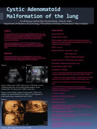
Modified classificationby Adzick • Macrocystic CCAMs single or multiple cysts>5mm in diameter, sonographically fluid-filled cysts 2. Microcystic CCAMs: more solid and bulky, <5mm in diameter, sonographically solid and homogenous (High mortality rate of microcystic CCAM due to large size and secondary sequale including mediastinal shift, pulmonary hypoplasia, polyhydroamnios and nonimmune hydrops)
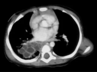
Prenatal Sonographic Findings of CCAM • Solid or cystic lung tumor without vascular flow • Type I or II: cystic or echolucent masses • ddx: CDH, cystic hygroma, brochogenic or enteric cyst, pericardial cyst • Type III: large hyperechogenic mass often associated with mediastinal shift and hydrops • Blood supply from the pulmonary artery and drain to pulmonary veins: bronchopulmoary sequestration and hybrid lesion: systemic feeding vessel
Postnatal History of CCAM • Postnatal Hx: quite variable, completely asymptomatic, mild respiratory complaints with recurrent infection, • 10% present after the first year of life • 80% present at birth with severe cardiorespiratory compromise due to severe pulmonary hypoplasia • 14% of CCAM result in stillbirth
Antenatal Natural History of CCAM • The outcome of fetuses with prenatal diagnosed CCAM: recently reported • Worst outcome: in the fetus with hydrops fetalis • Hydrops is usually seen in very large type III lesions, which causes mediastinal shift and vena caval obstruction • Rare survival of CCAM with hydrops, only exception those when fetal surgery has been performed
Antenatal Natural History of CCAM • Polyhydroamnios: esophageal obstruction from mediastinal shift, supported by the absence of fluid in the stomach • Mirror syndrome: hyperdynamic preeclamptic state that may be life-threatening, immediate delivery of the baby (the only treatment) • Spontaneous regression: 6 -11%, mechanism of regression (decompression of fluid from a CCAM into tracheobrochial tree, outgrowing its blood supply) • MacGillivary (1993): six cases of large CCAM with associated mediastinal shift • progressively decreased in size • all of the microcystic, or type III • none was associated with nonimmune hydrops fetalis
CCAM volume and CCAM volume ratioby Crombleholme in 1999 • predictor of the development of hydrops fetalis • CCAM volume : h x w x l x 0.52 in cm3 9 (ellipse) • CCAM volume ratio(CVR): CCAM volume/ head circumference • CCAM volume and CVR is significantly higher in fetus with hydrops fetalis • CVR <1.2 (cutoff value ): no hydrops fetalis
Large experience of prenatal diagnosed CCAM( San Francisco and Philadelphia) • 12-years retrospective study of two centers • 175 fetal lung lesions, 134 fetuses with CCAM • 14 pregnancies were terminated • 101 cases were managed expectantly • 13 women underwent open fetal surgery • 6 fetuses underwent thoracoamniotic shunt • Postnatal survival in non-hydrops fetalis: 100% • Postnatal or intrauterine mortality in hydrops fetalis(24 case) with expectant management: 100%
Management of pregnancy • Chromosomal abnormality: 0.7% • Color Doppler study to rule out BPD • Evidence of mediastinal shift and hydrops • Associated cardiac abnormality: fetal echocardiography • Prenatal consultation from a pediatric surgeons, neonatologist and pediatric cardiologist • If there are associated life-threatening anomaly: recommend not to continue pregnancy • If “mirror syndrome”, immediate delivery • Isolated CCAM without hydrops: close follow-up for fetal hydrops • All fetus with CCAM should be delivered at tertiary-care center
Fetal Intervention of CCAM • CCAM with hydrops after 32 wks: betamethasone Tx and early delivery for immediate postnatal resection • CCAM with hydrops before 32 wks: treatment in utero • thoracoamniotic shunt: Macrocystic lesion • fetal surgical resection: microcystic lesion • 13 cases of fetal surgical resection • definite resolution of fetal hydrops in 1-2 weeks • return mediastinal shift and impressive in-utero lung growth in 3 weeks • five fetal death: mirror syndrome(1), bradycardia(1), uncontrolled uterine contraction(1), intraoperative death(2)