Download
1 / 4
50 likes | 111 Views
The future laparoscopic technology includes threedimensional virtual reality and expands the scanning rate from 525 lines of resolution to 1,000 or 1,200 lines per frame and the quality of picture would be twice better than existing system.

E N D
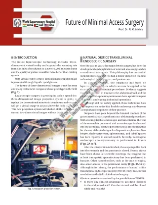
Future of M inim al Access Surgery Prof. Dr. R. K. Mishra INTRODUCTION The future laparoscopic technology includes three- dimensional virtual reality and expands the scanning rate from 525 lines of resolution to 1,000 or 1,200 lines per frame and the quality of picture would be twice better than existing system. With virtual reality, a three-dimensional computer image is presented through liquid crystal glasses. The future of three-dimensional images is not far away and many instrument companies have prototype in the field (Fig. 1). Laparoscopic surgery is growing in such a speed that three-dimensional image projection system is going to replace the conventional monitor in near future and surgeon will get a virtual image in air just above the body of patient. This new projection system will abolish all the limitation of current two-dimensional images without depth perception. NATURAL ORIFICE TRANSLUMINAL ENDOSCOPIC SURGERY Over the past 10 years, the major drive in surgery has been the development and application of minimal access approaches to traditional operations. This philosophy has crossed all surgical specialties and has had a major impact on training, technological developments, and patient care. In general surgery, the emphasis has been on laparoscopic techniques, which can now be applied to the majority of intra-abdominal procedures. Evidence suggests that the reduction in trauma to the abdominal wall and the physiology of the pneumoperitoneum have a positive impact on patients undergoing abdominal operations. Although still not widely applied, these techniques have put surgeons on notice that flexible endoscopy may become an important component of their practice. Surgeons have gone beyond the luminal confines of the gastrointestinal tract to perform intra-abdominal procedures. With existing flexible endoscopic instrumentation, the wall of the stomach is punctured and an endoscope is advanced into the peritoneal cavity to perform various procedures; thus far, the use of this technique for diagnostic exploration, liver biopsy, cholecystectomy, splenectomy, and tubal ligation has been reported in animal models. Recently, transvaginal endoscopic cholecystectomy is performed in France (Figs. 2A to F). After the intervention is finished, the scope is pulled back into the stomach and the puncture is closed. Several videos have been shown at scientific meetings that suggest that at least transgastric appendectomy has been performed in humans. Other natural orifices, such as the anus or vagina, also allow access to the peritoneal cavity (Figs. 3A to C). Although in its infancy, the performance of natural orifice transluminal endoscopic surgery (NOTES) may, thus, further revolutionize the field of abdominal surgery. Obvious questions are raised by the possibilities of NOTES: ■ Is there any clinical advantage to avoiding incisions in the abdominal wall? Can the visceral wall be closed safely and reliably? Fig. 1: Hologram projection system.
626 SECTION6: Miscellaneous A B C D E F Figs. 2A to F: Transvaginal cholecystectomy. ■ Does the flora of the natural orifice lead more to peritoneal infection? Who should perform these procedures and how should the individuals be trained in NOTES? The Society of American Gastrointestinal and Endoscopic Surgeons (SAGES) and gastroenterologists from the American Society for Gastrointestinal Endoscopy (ASGE) held a summit meeting to build a conceptual framework for the initial safe application of NOTES procedures. It was acknowledged that the development of NOTES further blurred the lines between the traditional boundaries of gastrointestinal surgeons and endoscopists. Several broad concepts and principles were agreed upon and will soon be published in detail in a whitepaper. surgeons and interventional endoscopists in an operating room. Clinical procedures should be done under Institutional Review Board Supervision and entered into a prospective outcomes database. Basic research is needed to assess the physiologic alterations caused by the visceral puncture and the bacteriology of the peritoneal cavity following transluminal interventions. Collaboration with industry is critical to the development of effective instruments allowing traction/countertraction and stable optical platforms as well as the means to control hemostasis, securely close the visceral wall, and perform suturing functions and gastrointestinal anastomoses. The roots of NOTES have been established, but work is still needed to refine techniques, verify safety, and document efficacy. With continued research, NOTES may prove to be a sound method with clinical benefit to patients. Appropriate scientific scrutiny, collaboration among gastrointestinal interventionalists, and effective means of training for these techniques will be critical on emerging technologies. ■ Safe Application of Natural Orifice Transluminal Endoscopic Surgery The concepts included the following: initially, NOTES should be performed by a team of experienced laparoscopic
627 CHAPTER49: Future of Minimal Access Surgery A B C Figs. 3A to C: Introduction of operative endoscope through the vaginal orifice. 3. Davis MC, Can DD, Pindrik J, Rocque BG, Johnston JM. Virtual interactive presence in global surgical education: international collaboration through augmented reality. World Neurosurg. 2016;86:103-11. 4. Gavaghan K, Oliveira-Santos T, Peterhans M, Reyes M, Kim H, Anderegg S, et al. Evaluation of a portable image overlay projector for the visualisation of surgical navigation data: phantom studies. Int J Comput Assist Radiol Surg. 2012;7:547-56. 5. Gavaghan KA, Peterhans M, Oliveira-Santos T, Weber S. A portable image overlay projection device for computer-aided open liver surgery. IEEE Trans Biomed Eng. 2011;58:1855-64. 6. Gonzalez SJ, Guo YH, Lee MC. Feasibility of augmented reality glasses for real-time, 3-dimensional (3D) intraoperative guidance. J Am Coll Surg. 2014;219:S64. 7. Ieiri S, Uemura M, Konishi K, Souzaki R, Nagao Y, Tsutsumi N, et al. Augmented reality navigation system for laparoscopic splenectomy in children based on preoperative CT image using optical tracking device. Pediatr Surg Int. 2012;28:341-6. 8. Inoue D, Cho B, Mori M, Kikkawa Y, Amano T, Nakamizo A, et al. Preliminary study on the clinical application of augmented reality neuronavigation. J Neurol Surg A Cent Eur Neurosurg. 2013;74:71-6. 9. Kang X, Azizian M, Wilson E, Wu K, Martin AD, Kane TD, et al. Stereoscopic augmented reality for laparoscopic surgery. Surg Endosc. 2014;28:2227-35. 10. Kenngott HG, Wagner M, Gondan M, Nickel F, Nolden M, Fetzer A, et al. Real-time image guidance in laparoscopic liver surgery: first clinical experience with a guidance system based on intraoperative CT imaging. Surg Endosc. 2014;28:933-40. 11. Kersten-Oertel M, Gerard I, Drouin S, Mok K, Sirhan D, Sinclair DS, et al. Augmented reality in neurovascular surgery: feasibility and first uses in the operating room. Int J Comput Assist Radiol Surg. 2015;10:1823-36. 12. Kocev B, Ritter F, Linsen L. Projector-based surgeon-computer interaction on deformable surfaces. Int J Comput Assist Radiol Surg. 2014;9:301-12. 13. Kranzfelder M, Bauer M, Magg M. Image-guided surgery. Endosk Heute. 2014;27:154-8. 14. Lahanas V, Loukas C, Smailis N, Georgiou E. A novel augmented reality simulator for skills assessment in minimal invasive surgery. Surg Endosc. 2015;29:2224-34. 15. LeBlanc F, Champagne BJ, Augestad KM, Neary PC, Senagore AJ, Ellis CN, et al. A comparison of human cadaver and augmented reality simulator models for straight laparoscopic colorectal skills acquisition training. J Am Coll Surg. 2010;211:250-5. 16. Lelos N, Campos P, Sugand K, Bailey C, Mirza K. Augmented reality dynamic holography for neurology. J Neurol Neurosurg Psychiatr. 2014;85:1. 17. Marescaux J, Diana M. Inventing the future of surgery. World J Surg. 2015;39:615-22. Fig. 4: Augmented reality in minimal access surgery. AUGMENTED REALITY IN MINIMAL ACCESS SURGERY Augmented reality is slowly emerging in minimal access surgery around the world to help the laparoscopic and robotic surgeons and the operating team (Fig. 4). We have seen surgeons wearing the Google glass and as technology evolves, both in terms of devices and software, we are seeing an extension in applications. Reaching a truly augmented surgeon through augmented reality will require few more years. For the time being, augmented reality and virtual reality are becoming more and more common in minimal access surgical training and in the preparation of laparoscopic surgery, letting the surgeon to practice on a virtual patient that mirrors, accurately, the real patient. The development of augmented reality devices allows minimal access surgeon to incorporate data visualization into diagnostic and treatment procedures to improve work efficiency, safety, and cost and to enhance surgical outcome. BIBLIOGRAPHY 1. Badiali G, Ferrari V, Cutolo F, Freschi C, Caramella D, Bianchi A, et al. Augmented reality as an aid in maxillofacial surgery: validation of a wearable system allowing maxillary repositioning. J Craniomaxillofac Surg. 2014;42:1970-6. 2. Callovini G, Sherkat S, Callovini G, Gazzeri R. Frameless nonstereotactic image-guided surgery of supratentorial lesions: introduction to a safe and inexpensive technique. J Neurol Surg A Cent Eur Neurosurg. 2014;75:365-70.
628 SECTION6: Miscellaneous 18. Nam WH, Kang DG, Lee D, Lee JY, Ra JB. Automatic registration between 3D intraoperative ultrasound and preoperative CT images of the liver based on robust edge matching. Phys Med Biol. 2012;57:69-91. 19. Okamoto T, Onda S, Matsumoto M, Gocho T, Futagawa Y, Fujioka S, et al. Utility of augmented reality system in hepatobiliary surgery. J Hepatobiliary Pancreat Sci. 2013;20:249-53. 20. Okamoto T, Onda S, Yanaga K, Suzuki N, Hattori A. Clinical application of navigation surgery using augmented reality in the abdominal field. Surg Today. 2015;45:397-406. 21. Pessaux P, Diana M, Soler L, Piardi T, Mutter D, Marescaux J. Robotic duodenopancreatectomy assisted with augmented reality and real-time fluorescence guidance. Surg Endosc. 2014;28:2493-8. 22. Pessaux P, Diana M, Soler L, Piardi T, Mutter D, Marescaux J. Towards cybernetic surgery: robotic and augmented reality- assisted liver segmentectomy. Langenbecks Arch Surg. 2015; 400:381-5. 23. Profeta AC, Schilling C, McGurk M. Augmented reality visualization in head and neck surgery: an overview of recent findings in sentinel node biopsy and future perspectives. Br J Oral Maxillofac Surg. 2016;54:694-6. 24. Shakur SF, Luciano CJ, Kania P, Roitberg BZ, Banerjee PP, Slavin KV, et al. Usefulness of a virtual reality percutaneous trigeminal rhizotomy simulator in neurosurgical training. Neurosurgery. 2015;11:420-5. 25. Shekhar R, Dandekar O, Bhat V, Philip M, Lei P, Godinez C, et al. Live augmented reality: a new visualization method for laparoscopic surgery using continuous volumetric computed tomography. Surg Endosc. 2010;24:1976-85. 26. Shenai MB, Dillavou M, Shum C, Ross D, Tubbs RS, Shih A, et al. Virtual interactive presence and augmented reality (VIPAR) for remote surgical assistance. Neurosurgery. 2011;68:200-7. 27. Sugimoto M, Yasuda H, Koda K, Suzuki M, Yamazaki M, Tezuka T, et al. Image overlay navigation by markerless surface registration in gastrointestinal, hepatobiliary and pancreatic surgery. J Hepatobiliary Pancreat Sci. 2010;17:629-36. 28. Tabrizi LB, Mahvash M. Augmented reality-guided neurosurgery: accuracy and intraoperative application of an image projection technique. J Neurosurg. 2015;123:206-11. 29. Tsutsumi N, Tomikawa M, Uemura M, Akahoshi T, Nagao Y, Konishi K, et al. Image-guided laparoscopic surgery in an open MRI operating theater. Surg Endosc. 2013;27:2178-84. 30. Vemuri AS, Wu JCH, Liu KC, Wu HS. Deformable three- dimensional model architecture for interactive augmented reality in minimally invasive surgery. Surg Endosc. 2012;26:3655-62. 31. Vera AM, Russo M, Mohsin A, Tsuda S. Augmented reality telementoring (ART) platform: a randomized controlled trial to assess the efficacy of a new surgical education technology. Surg Endosc. 2014;28:3467-72. 32. Watanabe E, Satoh M, Konno T, Hirai M, Yamaguchi T. The transvisible navigator: a see-through neuronavigation system using augmented reality. World Neurosurg. 2016;87:399-405. 33. Wen R, Chui CK, Ong SH, Lim KB, Chang SKY. Projection-based visual guidance for robot-aided RF needle insertion. Int J Comput Assist Radiol Surg. 2013;8:1015-25. 34. Yoshino M, Saito T, Kin T, Nakagawa D, Nakatomi H, Oyama H, et al. A microscopic optically tracking navigation system that uses high-resolution 3D computer graphics. Neurol Med Chir (Tokyo). 2015;55:674-9.