Download
1 / 33
360 likes | 755 Views
Application of otoacoustic emissions in the diagnosis of hearing loss. Bradley McPherson PhD Centre for Communication Disorders University of Hong Kong. Otoacoustic emissions. Otoacoustic emissions (OAE) are low intensity audio-frequency sounds

E N D
Application of otoacoustic emissions in the diagnosis of hearingloss Bradley McPherson PhD Centre for Communication Disorders University of Hong Kong
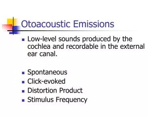
Otoacoustic emissions • Otoacoustic emissions (OAE) are low intensity audio-frequency sounds • Produced by the cochlea as part of the normal hearing process • Kemp (1978) English biophysicist discovered otoacoustic emission and published the first scientific description of transient-evoked otoacoustic emissions (TEOAEs) • In 1979 first work with distortion product otoacoustic emissions (DPOAEs) • TEOAEs are elicited by clicks or brief tonal stimuli, and DPOAEs are elicited by two simultaneously presented tones of slightly different frequency
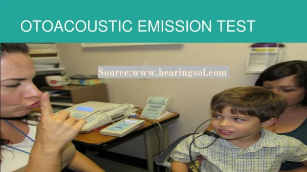
Uses of OAEs Screening using OAEs: • Neonatal hearing screening • Community-based hearing screening • School-based hearing screening • NIHL screening
Uses of OAEs Overall response = 20.4 dB SPL, 81% reproducibility Response at 1.5 kHz = 12 dB SPL, 94% reproducibility Response at 2.2 kHz = 11 dB SPL, 94% reproducibility Response at 3 kHz = 7 dB SPL, 84% reproducibility TEOAE result 3 day old neonate
Uses of OAEs Diagnostic uses of OAEs: • NIHL • Functional hearing loss • Retrocochlear hearing loss • Ménière’s disease • Sudden hearing loss
Case history 1 • Screening program for auditory neuropathy • January 2002 • Children attending school for deaf screened using otoscopy, tympanometry and TEOAE measures • KK tested positive in both ears • Bilateral TEOAEs present • Follow-up • Detailed case history from KK’s father and teacher • Diagnostic assessment in February 2002
KK’s history • 9;6 years, female • Initially diagnosed at 3 years with severe bilateral sensorineural hearing loss • Fitted with hearing aid for left ear at 3;4 years • At first full assessment also diagnosed with low muscle tone and balance disorder • Normal CT and MRI findings
KK’s history • Family history: mother and younger brother have hearing loss of unknown aetiology • Brother has mild loss but good speech and language development
KK’s history • Speech production: some unintelligible vowel-like utterances • Speech reception: very poor • Communication by sign at home and with classmates • Depends greatly on visual cues • Oticon BTE hearing aid in left ear of no significant benefit, even with FM system
OAE results • Confirmed TEOAE in right ear • Noted high frequency TEOAE in left ear • DPOAE results consistent with TEOAE results • DPOAE amplitude growth results also consistent with other findings
Other test results • Tympanometry: bilateral Type A • Acoustic reflexes: absent contralateral and ipsilateral • Speech recognition: 0% unaided in monaural and binaural conditions, 0% aided left
Other test results • ABR: no synchronous responses at 95 dB nHL in right or left ear • Cochlear microphonic present bilaterally • Middle latency response: absent right and left • Late evoked response: N1-P2 present bilaterally with ipsi- or contralateral stimulation
ABR in right ear Cochlear microphonic
Case history 2 • 26-year-old Chinese woman seen for an audiological evaluation in May 2003 • Main complaint was persistent hearing difficulties in situations with background noise, particularly in speech perception • She reported only occasional difficulty with speech understanding in quiet
Case history 2 • Recent pure-tone audiometric assessment had indicated normal hearing • Her reported communication disorder occurred after an episode of transient ischemic attack in November 2002 • Previously, she had been diagnosed with moyamoya disease following hospitalization for left temporoparietal intracerebral hematoma
Moyamoya disease • A rare condition - the progressive narrowing of the distal internal carotid arteries and proximal portions of the anterior and middle cerebral arteries • Most patients with moyamoya disease are children or adults in the third or fourth decades of life • Nearly two-thirds are female • In the later stages of MD ischemic episodes are common, with clinical symptoms such as impaired consciousness, focal motor symptoms, speech dysfunction, seizure and sensory disorders
Moyamoya disease • Moyamoya first identified in Japan in 1959 • ‘Moyamoya’ is Japanese for a ‘puff of smoke’ • Refers to the wispy cloud of fragile blood vessels seen on brain angiograms • These develop where normal vessels are blocked
Case history 2 • Clear, unoccluded ear canals; tympanic membranes were of normal appearance • Consistent responses to standard pure-tone audiometry • Normal hearing thresholds in the left ear and a very slight low frequency sensorineural hearing loss noted in the right ear • Tympanometry results were consistent with normal middle function in both ears. Acoustic reflexes with ipsilateral and contralateral stimuli were present in both the left and right ears
Case history 2 • Transient evoked OAEs showed clear responses - right and left ears • Over full range of frequencies measured (1000 Hz to 5000 Hz) • Normal OAEs with slightly reduced right ear thresholds indicated a need for further testing • Possible retrocochlear disorder
Case 2 TEOAEs Right ear Left ear
Case history 2 CAPD evaluation was also performed • Hearing in Noise Test - Cantonese version: Binaural 50% correct threshold for speech in quiet = 26 dBA. Binaural speech in noise composite threshold was -3.0 dBA. Both values outside the 99th percentile for normal listeners • Pitch Pattern Sequence: Results also abnormal 58% correct in the left ear and 70% in the right ear (norms = 99% correct score) • Random Gap Detection: All thresholds were markedly abnormal compared to clinical normative values (50 msec compared to 6.4 msec)
Case history 2 • CAPD test results were consistent with a diagnosis of central auditory processing disorder • Results were explained to the patient, who was relieved to learn that her auditory problems were ‘real’ despite earlier, pure-tone only evaluations having stated her hearing was normal • We were able to give the patient a clear understanding of the reason for her difficulties in situations of environmental noise
In conclusion • OAE information can be of great value in diagnostic audiology • Can help determine site of disorder • Quick, easy to perform and non-invasive • Should always be included in a full diagnostic assessment
Acknowledgements These case studies were only possible with the assistance of: • Dr Man-tak Leung, Teaching Fellow, HKU • Ms Lena Wong, Assistant Professor, HKU • Mr Kevin Yuen, Research student, CUHK • Ms Juvy Lee & Ms Tempo Tang, Research students, HKU