Endotracheal Intubation
670 likes | 2.38k Views
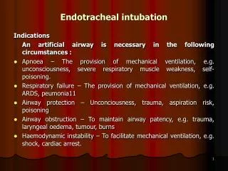
Endotracheal Intubation. Advantages of Intubation. A cuffed endotracheal tube protects the airway from aspiration Access is gained to the tracheobronchial tree for the suctioning of secretions Ventilations via an entotracheal tube do not cause gastric distention

Endotracheal Intubation
E N D
Presentation Transcript
Advantages of Intubation • A cuffed endotracheal tube protects the airway from aspiration • Access is gained to the tracheobronchial tree for the suctioning of secretions • Ventilations via an entotracheal tube do not cause gastric distention • Maintains a patent’s airway and assists in avoiding further obstruction • Enables delivery of certain medications
Indications • For supporting ventilation in patient with :- • Upper airway obstruction • Respiratory failure • Loss of conciousness • For supporting ventilation during general anesthesia. • Patients atrisk of pulmonary aspiration • Difficult mask ventilation • Any patient in imminent danger of upper airway obstruction (e.g. Burns of the upper airways). • Cardiac arrest
Contraindications • A patient with an intact gag reflex • Patients likely to react with laryngospasm (i.e. children with epiglottitis) • Cervical spine injury
Condition that associated with difficult intubation • Congenital anomalies Down’s syndrome • Infection in airway Retropharyngeal abscess, Epiglottitis • Tumor in oral cavity or larynx • Enlarge thyroid gland trachea shift to lateral or compressedtracheal lumen • Maxillofacial ,cervical or laryngeal trauma • Temperomandibular joint dysfunction • Burn scar at face and neck • Morbid obesity
Air way assessment 1.Mallampati classification • This test is performed with the patient in the sitting position, head in a neutral position, the mouth wide open and the tongue protruding to its maximum • Class I: Visualization of the soft palate, uvula, anterior and the posterior pillars. • Class II: Visualization of the soft palate and uvula. • Class III: Visualization of soft palate and base of uvula. • Class IV: Only hard palate is visible. Soft palate is not visible at all.
Soft palate Uvula Class III, IV difficult to intubate
2. Interincisorgab: • Normal >4.5 cm (3 fingers)
3) Thyromental distance : more than 6 cms • 4) Flexion and extension of neck
5. Laryngoscopic view • Grade 3,4 risk for difficult intubation!
Preparing the procedure... Essentials that must be present to ensure a safeintubation!.. They can be remembered by the mnemonic SALT • Suction. This is extremely important. Often patients will have secretions in the pharynx, making visualization of the vocal cords difficult. • Airway. the oral airway is a device that lifts the tongue off the posterior pharynx, often making it easier to mask ventilate a patient. Also a source of O2 with a delivery mechanism (ambu-bag and mask) must be available. • Laryngoscope. This is vital to placing an endotracheal tube. • Tube. Endotracheal tubes come in many sizes. In the average adult a size 7.0 or 8.0 endotracheal tube
Instruments used... • Self-refilling bag-valve combination (eg, Ambu bag), tubing, and oxygen source. • Plaster or tube holder . • Introducer (stylets or Magill forceps). • Laryngoscope • Suction apparatus • Syringe, 10-mL, to inflate the cuff. • Mucosal anesthetics (eg, 2% lidocaine) • Water-soluble sterile lubricant. • Gloves. • Pulse oximeter • Stethoscope
Oral airway Nasal airway • Oropharyngeal or nasopharyngeal airway
Miller blade Macintosh blade LARYNGOSCOPIC BLADE Macintosh (curved) and Miller (straight) blade Adult : Macintosh blade, small children : Miller blade
Endotracheal tube • Size of endotracheal tube : internal diameter (ID) • Male: ID 8.0 mms . Female : ID 7.5 mms • New born - 3 months : ID 3.0 mms • 3-9 months : ID 3.5 mms • 9-18 months : ID 4.0 mms • 2- 6 yrs : ID = (Age/3) + 3.5 • > 6 yrs : ID = (Age/4) + 4.5
Depth of endotracheal tube : Midtrachea or below vocal cord ~ 2 cms Adult: Male = 23 cms ,Female = 21 cms Children: endotracheal tube = (Age/2) + 12 (cm)
Tecnique: Sniffing position Flexion at lower cervical spine Extension at atlanto-occipital joint
Tecnique Make sure that all materials are assembled and close at hand Make sure that the balloon inflates Check the laryngoscope and blade for proper fit, and make sure that the light works Anesthetize the mucosa of the oropharynx, and upper airway withlidocaine 2%, if time permits and the patient is awake. Hyperventilate the patient with 100% oxygen for 1 minute prior to intubation attempt Place the patient in the sniffing position.
Open the patient's mouth with the right hand, and remove any dentures. • Grasp the laryngoscope in the left hand • Spread the patient's lips, and insert the blade between the teeth, being careful not to break a tooth. • Pass the blade to the right of the tongue, and advance the blade into the hypopharynx, pushing the tongue to the left. • Lift the laryngoscope upward and forward, without changing the angle of the blade, to expose the vocal cords.
Takethe endotracheal tubein the right hand and starts inserting it through the mouth opening. The tube is inserted through the cords to the point that the cuff rests just below the cords (between 21-23 mark on the tube) Holding the tube firmly in place, quickly remove the laryngoscope Remove the stylet from the endotracheal tube Finally, the cuff is inflated with 5-10 ml of air Ventilate the patient Observing the chest rise and fall with each ventilation
Listens for breathing sounds to ensure correct placement of the tube (in stomach and chest) If no breath sounds and there is bubble sound in stomach (it is in stomach) remove the tube and ventilate the patient and start all over again If the tube is advanced too far, it will get into the right bronchus and only the right lung is ventilated. If this occurs deflate the cuff with draw 2-3 cm and re-inflate the cuff and listen again Attach the tube to the patient and to the ventilating apparatus
Complication of endotracheal intubation • 1) During intubation • 2) During remained intubation • 3) During extubation • 4) After extubation
1) During intubation • Laryngeal edema • Arytenoid dislocation hoarseness • Increased intracranial pressure • Spinal cord trauma in cervical spine injury • Esophageal intubation • Trauma to lip, tongue or teeth • Hypertension and tachycardia or arrhythmia • Pulmonary aspiration • Laryngospasm • Bronchospasm
2) During remained intubation • Obstruction from secretion or overinflation of cuff • Accidental extubation or endobronchial intubation • Disconnection from breathing circuit • Lib or nasal ulcer in case with prolong period of intubation
3) DuringExtubation • Laryngospasm • Pulmonary aspiration • Edema of upper airway
4)After Extubation • Sore throat • Hoarseness • Tracheal stenosis (Prolong intubation) • Laryngeal granuloma