Download
1 / 15
240 likes | 1.21k Views
Explore the visual representation of human body structures through cross-sectioned cadaveric specimens. Learn how this aids in CT interpretation and appreciation for anatomical relationships.

E N D
Cross section anatomy of ABDOMEN Drgauthamkamble
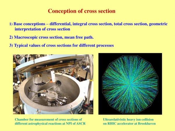
The study of Cross-Sectional Anatomy serves two purposes; • as a visual medium to facilitate an understanding of the structural organization of the human body • as preparation for the study of computed tomography (CT).
A study of human anatomy from cross-sectioned cadaveric specimens provides a visually unique representation of the positions, sizes, shapes, and relationships of structures • this in turn facilitates an appreciation for the three-dimensional organization of the body. • Cross-sectioned cadaveric images permit observation of intact anatomical relationships in specimens in a highly unique manner. • Within the clinical sciences a study of cross-sectioned cadaveric specimens facilitates interpretation of CT radiology since CT scans are typically performed in the cross-sectional plane.
CT scan or Computed Tomography uses a combination of X-rays and computer technology to produce detailed, cross-sectional image of the body. Magnetic resonance imaging (MRI) of the body uses a powerful magnetic field, radio waves and a computer to produce detailed pictures of the inside of the body.
Transpyloric plane • Imaginary transverse plane • Anteriorly passes through the tips of 9th costal cartilage • Posteriorly through the lower border of 1st lumbar vertebra • Midway between suprasternal notch and pubic symphysis • Roughly a hand s breadth below xiphisternal joint
Pylorus of stomach • Hila of kidneys • Fundus of gall bladder • Neck of pancreas • Origin of coeliac trunk • Origin of superior mesenteric artery
Subcostal plane • Transverse plane passing through the lowest part of costal margin • Lowest costal margin formed by 10 th costal cartilage • Passes through junction of 2nd and 3rd lumbar vertebra