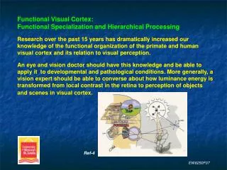
EW8250F07
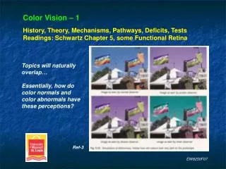
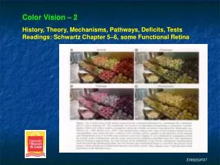
Color Vision – 2 History, Theory, Mechanisms, Pathways, Deficits, Tests Readings: Schwartz Chapter 5–6, some Functional Retina. Ref-3. EW8250F07. EW8250F07.

EW8250F07
E N D
Presentation Transcript
Color Vision – 2History, Theory, Mechanisms, Pathways, Deficits, TestsReadings: Schwartz Chapter 5–6, some Functional Retina Ref-3 EW8250F07
Cone PhotopigmentsEach cone contains millions of molecules of photopigment.The photopigment in each cone is the same.Generally there are 3 classes of photopigments, thus 3 cone types.Each cone type has a particular spectral sensitivity:range of wavelength absorption and peak absorption efficiency.Short Wavelength Sensitive (SWS) = S-cones, peak absorption ~425 nmMedium Wavelength Sensitive (MWS) = M-cones, peak absorption ~530 nmLonger Wavelength Sensitive (LWS) = L-cones, peak absorption ~560 nmPreviously (incorrectly) called blue (SWS), green (MWS) and red (LWS) cones.Because each does not respectively signal only blue, green and red light.Also, peak absorption for LWS cones is for yellow-green and not red. EW8250F07
1.2 1.0 0.8 Relative sensitivity 0.6 0.4 0.2 0.0 400 425 450 475 500 525 550 575 600 625 650 675 700 Wavelength (nm) The 3 general photopigment, or cone, classes have broadly overlapping ranges of spectral sensitivity (absorption). Cut-offs to note: ~ 525 nm ~ 650 nm EW8250F07
Absolute Sensitivity of the 3 Photopigments (Cones)L-cone > M-cone >> S-coneCone envelope (photopic sensitivity) < rod (scotopic sensitivity) Ref-2 EW8250F07
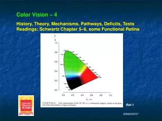
Spectral sensitivity is measured via microspectrophotometry(in vitro suction electrodes, monochromatic light stimulation)and visual psychophysics (various color matching methods).Data from different techniques and species match well. In vitro primate data (human data matches): Note the S-cone equal sensitivity due to no filtering by media and macular pigment. Filters protect against ultraviolet light damage and glare. Note the convergence of sensitivity for M- and L-cones at low wave- lengths. This does not occur in psychophysics (next slide). Ref-1 EW8250F07
Spectral sensitivity is measured via microspectrophotometry(in vitro suction electrodes, monochromatic light stimulation)and visual psychophysics (color matching, adaptation, etc.).Data from different techniques and species match well. Human data – psychophysics. Differences exist between normals and are due to: Filtering differences (lens, media and macular pigment), and photopigment variations (polymorphisms). Ref-1 EW8250F07
Opsin determines the absorption spectrumS-, M- and L-cones have a different opsin.Photopigment molecule: chromophore (11-cis-retinal) + opsin.Chromophore: identical for all photopigments; absorbs quanta.Opsin: transmembrane amino acid chain in the outer segment.Chromophore is embedded within and attached to the chain protein portion (i.e. the opsin) of the photopigment molecule. Absorption: opsin = 300 nm, retinal = 380 nm, linked together = broad range (dependent on the opsin amino acid sequence).This change in absorption of the chromophore when bound toa particular opsin is called the opsin-shift. EW8250F07
Opsins Are Encoded By Separate GenesRhodopsin (rod opsin) gene: on chromosome 3S-cone photopigment gene: on chromosome 7; single copyM- and L-cone photopigments genes: on the X chromosome in a head-to-tail tandem arrayNumber of genes in general:one copy for the L-cone pigment,multiple copies (1–5, mean 2) for the M-cone photopigment.Multiple copies does not appear to affect color vision. EW8250F07
Homology (identical DNA) exists between the gene of rhodopsin and those of the cone photopigments. This suggests a common ancestor of all 4 photopigments. The strongest homology is between rhodopsin and S-cones.M- and L-cone homology (identical DNA) = 98% = extremely strong.Suggests that both evolved recently.M- and L-cone to S-cone homology = 40% = not strong.Suggests that the S-cone evolved earlier. Pink = amino acids unequal EW8250F07
Ripe fruit in green background:evolutionary argument forthe development (i.e. need)of the L-cone photopigment. Area V8 is another higher-order color center. EW8250F07
Opsin Gene VariantsTypically only involves the M- and L-conesPolymorphic variants: cause slight shifts in color vision in normalsExamples: L-cone opsin at codon 18 (position in the opsin chain) contains either alanine or seranine. The seranine variant peak spectral sensitivity is shifted 3 nm toward the red relative to the alanine variant. For two corresponding M-cone variants the spectral sensitivity shift is up to 5 nm.Hybrid variants: cause abnormal color vision EW8250F07
Hybrid VariantsAre responsible for particular color deficiencies.Produced by fusion genes which contain partial coding sequence of both the M- and L-cone pigment genes (intragenetic crossover).Parts of the photopigment gene of the M-cone and L-cone fuse because they lie head-to-tail tandem array on the X chromosome.There are a range of possible hybrid photopigment genes.Amino acid substitutions shift the peak spectral sensitivity and can decrease the photopigment density, thus reducing its absorption (quantum efficiency).Hybrid pigments may be less stable. EW8250F07
Hybrid Gene FormationDuring meiosis – X chromosome alignment and exchange of information – unequal homologous recombination occurs. L-cone gene M-cone gene Normal meiosis. Normal expression of photopigments. No M- or L-cone gene. Deuteranope. Multiple M-cone genes without color anomaly Hybrid genes: normal, missing M or L, or partial M or L. Partial = anomalous; possible mechanism of anomalous trichromacy. Intergenetic crossover Intragenetic crossover Ref-2 EW8250F07
Very useful for learning and recall of red-green dichromat and anomalous trichromat deficits.Genes lie on different chromosomes for S-cone photopigment (7) and rhodopsin (3). Ref-2 EW8250F07
Prevalence and inheritance of color vision deficits. Inherited red-green deficits are transmitted as X-linked recessive. Inherited blue deficits are transmitted as autosomal dominant. EW8250F07
Examples of transmissionof X-linked, recessive,red-green deficitsTransmission is from motherto son so males with deficits have mothers who are carriers or have deficits.Transmission is recessive so a female must be homozygous to express the deficit. A femalewho is heterozygous is only acarrier (but may show shifts incolor vision in laboratory tests).Set-up the 2x2 table to determine or confirm answers to transmission questions. Ref-2 EW8250F07
What happens to the spectral luminosity function in dichromacy and anomalous trichromacy? R/G Anomalous Trichromacy Post-shift is not shown here but expect that sensitivity to red (≥ 650 nm) is maintained in deuteranomaly but is lost in protanomaly. Ref-2 EW8250F07
What happens to the spectral luminosity function in dichromacy and anomalous trichromacy? R/G Dichromacy Relative to the normal function, note the large shift for protanopia and very small shift for deuteranopia. Would you advise your patient to enter a health profession such as optometry if you diagnosed protanopia or severe protanomaly? Ref-1 EW8250F07
What happens to the spectralluminosity function in dichromacyand anomalous trichromacy?Fine schematic for anomaloustrichromats but…Not as accurate a schematicfor dichromats (simply removesone function) – use Schwartz. Ref-1 EW8250F07
References1. Norton, T., Corliss, D., & Bailey, J., 2002. The Psychophysical Measurement of Visual Function. Butterworth Heinemann.2. Schwartz, S., 2004. Visual Perception: A Clinical Introduction, 3rd Ed. McGraw-Hill.3. Snowden, R., Thompson, P., & Troscianko, T. 2006. Basic Vision: An Introduction to Visual Perception. Oxford.4. Palmer, S., 1999. Vision Science – Photons to Phenomenology. Massachusetts Institute of Technology.5. Rodieck, R. 1998. The First Steps In Seeing. Sinauer.6. Daw, N. 2006. Visual Development, 2nd Ed. Springer.7. Kaufman, P., & Alm, A. (Eds.). 2003. Adler’s Physiology of the Eye, 10th Ed. Mosby.8. Hubel, D. (1995). Eye, Brain and Vision. Scientific American Library.9. Oyster, C. 1999. The Human Eye: Structure and Function. Sinauer. EW8250F07