Download
1 / 24
240 likes | 353 Views
In the past 15 years, research has expanded our understanding of the functional organization of the primate and human visual cortex, particularly concerning visual perception. Eye and vision professionals must grasp how luminance energy transforms from local retinal contrast to the perception of objects and scenes. The identification of distinct visual areas highlights functional specialization—processing specific visual aspects like color and motion—and hierarchical processing, where local information transitions into abstract perceptions through ventral and dorsal pathways.

E N D
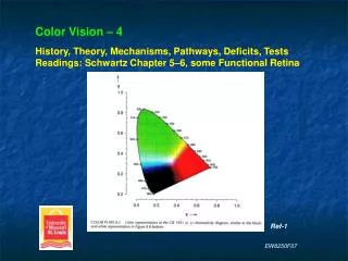
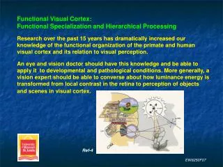
Functional Visual Cortex:Functional Specialization and Hierarchical ProcessingResearch over the past 15 years has dramatically increased our knowledge of the functional organization of the primate and human visual cortex and its relation to visual perception.An eye and vision doctor should have this knowledge and be able to apply it to developmental and pathological conditions. More generally, a vision expert should be able to converse about how luminance energy is transformed from local contrast in the retina to perception of objects and scenes in visual cortex. Ref-4 EW8250F07
A major accomplishment in visual neuroscience has been the identification of more than a dozen distinct visual areas.This has been gained via non-invasive methods (mainly FMRI) in humans and cell physiology in primates.Why are there so many visual areas? Two main explanations are: functional specialization and hierarchical processing.Functional specialization proposes that neural pathways and areas exist that process information about specific aspects of the visual scene: color, depth, motion and objects.Hierarchical processing proposes that a stage-wise process occurs in which local information is gradually transformed into abstract, holistic and multi-model information, culminating in perception. This process would occur separately for the ventral (‘what’) and dorsal (‘where’) pathways. EW8250F07
Background. Retinotopic maps obtained using a rotating wedge (pictures and colors optimally stimulate) reveal multiple horizontal and vertical meridian representations arranged in approximately parallel bands along the cortical surface. The meridians alternate and define borders between nearly mirror-symmetric retinotopic areas. These are called polar maps. Ref-1 EW8250F07
Polar Maps of Visual Areas EW8250F07
Background. Perpendicular to the meridian bands are eccentricity bands. These maps are obtained via an expanding ring and show a gradient as the foveola and fovea is greatly represented relative to the periphery. Ring radius = 1.5, 3–6, and 12–24 degrees. Importantly, retinotopic maps do exist in higher- order cortex, with polar maps declining faster than eccentricity maps. So visual cortex cannot simply be divided into retinotopic versus non- retinotopic areas. Ref-1 EW8250F07
Eccentricity Maps of Visual Areas(C and D) Ref-4 EW8250F07
Retinotopic maps are continuous through higher-order cortex. Visual areas are shown as parallel, nearly mirror-symmetric bands.This is important because the ability to identify specific areas then allows testing their function. V7 MT V4 V8 Ref-1 EW8250F07
Evidence for Functional SpecializationFunctional specialization proposes that neural pathways and areas exist that process information about specific aspects of the visual scene: color, motion, depth and objects.1. V4/V8 ~dedicated color center. Controversy will last awhile.2. MT/MST/STS ~dedicated motion center. No controversy.3. V1/V2/V3/VP/V3a/MT stream for depth. Relatively few studies.4. Ventral and dorsal paths, anterior to early cortex, for objects. Three main areas: LOC (lateral occipital complex), VOT (ventral occipito-temporal, and dorsal foci. Perhaps the strongest evidence. EW8250F07
Typical stimuli for faces, places and objects test. Ref-2 EW8250F07
Object selective regions shows functional specialization.LOC = lateral occipital complex = object-selective areas, including faces.LO = lateral occipital = dorsal region of the LOCpFus = posterior fusiform = ventral region of the LOC. Ref-1 EW8250F07
Object Selective Areas. FFA = fusiform face area, PPA = parahippocampal place area, both part of the VOT (ventral occipito-temporal). Note the locations relative to V1–V3. EW8250F07
Object Selective Areas. Anterior regions of VOT (ventral occipital temporal) tuned to faces (left), objects (center) and places (right). STS = superior temporal sulcus. Encodes biological motion! Face areas are more central than place areas. Ref-1 EW8250F07
Functional Specialization: V1 and V4 respond to all stimuli whereas areas LO and FFA respond differentially to detection, identification and no detection of faces. So higher-order areas involve perception. Ref-1 EW8250F07
Functional Specialization: Areas responsive to categories of objects. This meta-analysis of 18 studies shows strong consistency with regard to the location of activity. Ref-1 EW8250F07
Evidence for Hierarchical ProcessingHierarchical processing proposes that a stage-wise process occurs in which local information is gradually transformed into abstract, holistic and multi-model information, culminating in perception. This process would occur separately for the ventral (‘what’) and dorsal (‘where’) pathways. 1. Changes in neuron tuning and receptive field size.2. Changes in response to contrast.3. Changes in response to object adaptation.4. Changes in response to image scrambling.These studies have often involved mostly the ventral stream. EW8250F07
Evidence for Hierarchical Processing1. Changes in neuron tuning and receptive field size. V1 to LO receptive fields increase in size at every level. V1 neuron tuning is largely to spatio-temporal luminance. V2 neuron tuning includes illusory contours. V4 neuron tuning includes specific colors and forms. LO neuron tuning includes specific objects. 2. Changes in response to contrast. Sensitivity to contrast changes decreases from V1 to LO. LO response is essentially contrast invariant above 10%. MT/MST becomes contrast invariant at similar low levels. Ref-1 EW8250F07
Evidence for Hierarchical Processing3. Changes in response to object adaptation. Repeat viewing of an object produces reduced neural activity in LO and VOT but not in V1 and V2.4. Changes in response to image scrambling. Viewing gradually scrambled images of objects produces decreasing neural activity in LO and VOT but not in V1 and V2. Ref-1 EW8250F07
Evidence for Hierarchical ProcessingY-axis = FMRI response ratio = moving / staticX-axis = FMRI response ratio = preferred (tuned) stimulus / non-preferredFont size = degree of retinotopyPlot shows progressive tuning of neurons with higher-order areas.Also the local to globalprocessing with higher-order areas; indicatedby smaller retinotopy. Ref-1 EW8250F07
Evidence for Hierarchical ProcessingFrom the plot:V1–V3 are not finely tuned for motion or objects.V3a and V4 show more tuning to objects and motion.MT is tuned to motion much greater than to objects.PPA, FFA and LO showtuning to objects and not motion. Ref-1 EW8250F07
Evidence for Hierarchical Processing(a) FMRI response to increasing contrast becomes essentially invariant above 10%for V4 and LO but notfor area V1.(b) FMRI response to increasing stimulus size is essentially invariant for LO, more tuned for V4, and most sensitive for area V1. Ref-1 EW8250F07
Functional Specialization and Hierarchical ProcessingRight hemisphere.U = upper, D = down (lower).Note the orientation of the axes for specialization and hierarchy.Hierarchy: staircase showing the increase in abstract and holistic information from V1 to higher-order areas.Specialization: colors show the central (C) and peripheral (P) visual fields which changes in higher-order areas to specialized objects (O) and faces (F) areas centrally, and places (PL) peripherally. Ref-1 EW8250F07
Central-peripheral specializationextends from V1 to higher-order areas. Thus objects and faces not coincidently overlap with central representations and buildings and scenes overlap with peripheral representations.Hierarchical processingextends from V1 to higher-order areas. This correlates to the more anterior areas processing even more abstract, holistic and multi-modal representations.Therefore the two explanations for having many visual areasare likely a single explanation –both exist and function as one. Ref-1 EW8250F07
To appreciate vision…involves knowledge of object space, retinal image and cortex. Ref-4 Ref-1 EW8250F07
References1. Grill-Spector, K., & Malach, R. (2004). The human visual cortex. Annual Review of Neuroscience, 27.2. Chalupa, L., & Werner, J. (Eds.) (2004). The visual neurosciences. Bradford, MIT.3. Farah, M. (2000).The cognitive neuroscience of vision. Blackwell Publishers, UK.4. Snowden, R., Thompson, P., & Troscianko, T. (2006). Basic Vision: An Introduction to Visual Perception. Oxford.5. Wandell, B. (1995). Foundations of vision. Sinauer Associates, MA.6. Bruce, V., Green, P., & Georgeson, M. (2003). Visual perception: physiology, psychology and ecology. 4th Ed. Psychology Press, NY. EW8250F07