Download
1 / 46
520 likes | 1.35k Views
MALIGNANT MASSES IN BREAST ULTRASOUND. Dr. Mona Rozin Director of Breast Imaging Assuta Medical Centers. Usual Breast Cancers. DCIS IDC Medullary Mucinous Tubular Papillary ILC. I. DCIS. Precursor of IDC Become IDC after 5-8 yrs

E N D
MALIGNANT MASSES IN BREAST ULTRASOUND Dr. Mona Rozin Director of Breast Imaging Assuta Medical Centers mrozin,md
Usual Breast Cancers • DCIS • IDC • Medullary • Mucinous • Tubular • Papillary • ILC mrozin,md
I. DCIS • Precursor of IDC • Become IDC after 5-8 yrs • “dirty borders” – 30% will have a recurrence and 50% of these will be IDC • 30% of mammographically detected cancer in women aged 40-49 yrs. mrozin,md
DCIS - Prognosis • Most important feature is GRADE • Necrosis brings calcifications • Most are multifocal and 12% multicentric mrozin,md
DCIS - Mammo • Calcifications alone 70% • Calcifications in a mass or density 15% • Mass without calcifications 15% • Calcifications are “tip of the iceberg” especially in Low Grade DCIS mrozin,md
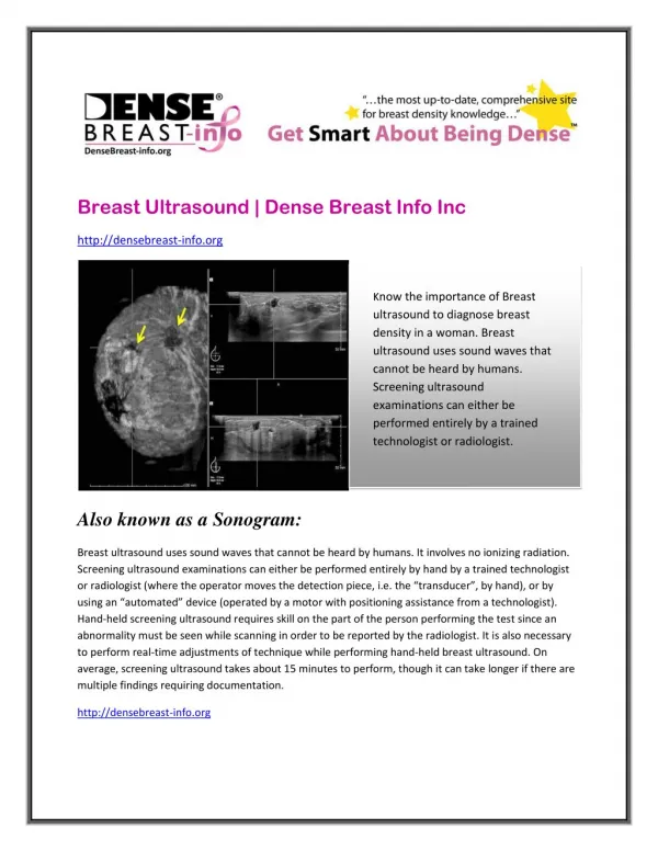
DCIS - Ultrasound • Mass – most common finding may see any of the entire BIRADS descriptors • Calcifications – may be seen with high frequency transducers increased vascularity on Doppler may have associated mass • Check mammo mrozin,md
DCIS - Ultrasound • May be mixed solid and cystic • Clustered “microcysts” –rare but these are usually vascular (Doppler), with thick walls and irregular shape & size • Abnormal size and branching of ducts THE GREAT PRETENDER mrozin,md
perpendicular parallel mrozin,md
microcysts min perpendicular max min parallel max mrozin,md
IDC DCIS mrozin,md
RT. normal LT. necrosis & calcs, desmoplasia, inflammatory infiltrates mrozin,md
2 1 adenosis with microcysts 5 DCIS with calcs 4 3 PURE DCIS DCIS microcysts mrozin,md
II. IDC • 75-80% of all CA • Host reaction: desmoplastic, inflammatory • Necrois: 30% - may be liquid, hemorrhagic, or fibrotic !!!!!!! • Intraductal component – 85% • Multifocal – 25-50% muticentric – 15-20% and bilateral – 5-8% • Invasion of Cooper’s ligaments – skin dimpling • Lymph node metastasis mrozin,md
I II III echogenic rim III II I branch size mrozin,md
I III Size of microlobulations mrozin,md
necrosis with central scar cystic necrosis mrozin,md
duct extension calcifications mrozin,md
III. Medullary CA • 5% of all CA, younger than 40yr, rapidly growing – therefore can be 3-4 cm, DCIS • US: hypo, circumscribed, enhancement !!!! mrozin,md
echogenic halo microloblulation mrozin,md
IV. Mucinous CA • 2% of all CA, older than 60 yrs, tumor cell mixed with mucin, DCIS – 75% • US: iso - “salt and pepper”, circumscribed, normal to enhanced transmission mrozin,md
taller than wide enhancement mrozin,md
V. Tubular • 2% of all CA, 40-50 yrs, DCIS - 75% • US: spiculation, thick echogenic rim, hypo, shadowing mrozin,md
VI. Papillary CA • 1-2% of all CA, 60-70 yrs, 50% arise in intraductal papillomas, bleeding nipple – 20-35%, may be intracystic, mixed with DCIS, abnormal ducts with enlarged TDLU mrozin,md
VII. ILC • 5-15% of all CA • “Indian files” on pathology • May not be detected on mammo, us, or physical exam (vague thickening) • Therefore may be found only when large! • Multifocal, multicentric, bilateral – MRI ! • Unusual metastasis – peritoneum, stomach, ovaries, meninges mrozin,md
ILC - Mammo • High false negative • May have no desmoplastic reaction • Isodense with few if any ca++ • May be seen on 1 view only (CC) • Spiculated mass, arch. distortion, asymmetric density, nipple & skin retraction • “Shrinking breast” !!!! mrozin,md
ILC - Ultrasound • Irregular shape 88% • Ill defined margins 94% • Shadowing 84% • Hypoechoic 92% • Wider than tall 77% • May also be iso or hyperechoic • Up to 10% not seen on US !!! • US is INVALUABLE in the staging of ILC mrozin,md
ILC Smaller left breast Indian files Diffuse enhancement Panoramic view mrozin,md
ILC Asymmetry mrozin,md
ILC Left shrunken breast mrozin,md
ILC “Golden Gate Sign” mrozin,md
Other Malignancies • Lymphoma • Metastasis • Sarcomas - angio SA- fibro SA - carcino SA • Inflammatory mrozin,md
Primary Lymphoma • 0.1 – 0.5% of all breast masses, 50-60 yrs, LYM:IDC 1:1000 • Mammo: increased density with skin thickening, large mass, path. ax. nodes-50% • US: diffuse hypoechogenicity, edema, dilated lymph channels, skin thickening mrozin,md
Primary Lymphoma mrozin,md
Metastasis to the Breast • Rare • Melanoma, lung, ovary, thyroid • Mammo/US: multiple bilateral nodules circumscribed or ill defined margins hypoechoic NO desmoplastic reation NO calcifications mrozin,md
Angio SA • 33% with negative mammo • 33% non-calcified mass • Purplish – blue skin discoloration • Hematogenic metastasis to lungs mrozin,md
Carcinosarcoma = Metaplastic CA • Rarest primary malignancy of the breast – less than 0.1% • Contains BOTH carcinomatous and sarcomatous components • Large and palpable – up to 5 cm, poorprognosis ! • Mammo: oval mass with circumscribed or ill defined margins, +/- calcifications • US: oval, ill defined margins, hypo, very vascular, enhancement, mixed solid/cystic mrozin,md
Carcinosarcoma = Metaplastic CA mrozin,md
2001 2002 mrozin,md
mag mrozin,md
Inflammatory CA • 1% of all CA, pregnancy & lactation • Erythema, warmth, edema in more than 1/3 of the breast associated with a high grade IDC, poor prognosis – 55% survival with chemo and rad Tx, 80% have tumor emboli in skin lymphatics • Mammo: edema, thick skin, thick C.L. • U/S: as in mammo, may need 5 MHz to penetrate and see hypoechoic mass mrozin,md
Inflammatory CA mrozin,md
Thank You ! mrozin,md