Normal Ultrasound Protocol Breast
410 likes | 1.33k Views
Normal Ultrasound Protocol Breast. Evidence 3. The following is a series of images that parallel the protocol as discussed in Evidence 2 All images are of the same patient.

Normal Ultrasound Protocol Breast
E N D
Presentation Transcript
Normal Ultrasound Protocol Breast Evidence 3
The following is a series of images that parallel the protocol as discussed in Evidence 2 • All images are of the same patient. • The patient (30 y/o) presented with a lump in the LEFT breast. The lump was palpated by the patient 4 days prior however it was not identified by the referring GP. The patient indicated and described a small lump in 12 o’clock position. • The ultrasound requested both breast be examined. • The patient did not indicate any concerns on the right breast. • The examination was performed on a Toshiba Aplio 500 using a 12Mhz multiple frequency .
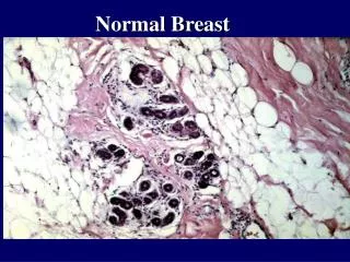
Normal breast • 12 images of the breast at each hour of the clock • Images are annotated with the side and body marker to show probe position
Images 1-4: Normal images in the o’clock position after appropriate scouting. All images display normal breast tissue.
Images 5-8: Normal images in the o’clock position after appropriate scouting. All images display normal breast tissue.
Images 9-12: Normal images in the o’clock position after appropriate scouting. All images display normal breast tissue.
Nipple • Longitudinal and transverse image of the nipple
Images 13-14: Longitudinal and Transverse images of the nipple respectively.
Axilla • Images of any lymph nodes in the short axis with cortical measurements of reactive or suspicious nodes
Image 15: Transverse image of the Axilla. A normal appearing lymph node is seen. The short axis and cortical thickness are measured and are within normal limits.
Image 16: Transverse image of the Axilla. The same lymph node is seen. Low scale Colour Doppler does not show any increase in vascularity.
Image 17: Longitudinal Image of the axilla. No suspicious lymph nodes seen
Pathology • Must be measured in two planes at 90 degrees unless a simple cyst
Images 18 - 19: Two images of the pathology at 90 degrees displaying measurement. Note the change in body marker to indicate position
Pathology • Colour imaging should be used to assess any vascularity
Image 20: Doppler image indicated no increased vascularity within the lesion.
Image 21: Additional image identifying location and position of pathology.
Pathology • Images of ROI must be taken and annotated Note: The following images are of the left breast
Images 22-25: Specific images of the area of interest as indicated by the patient. In this region normal breast tissue is seen.
Personal Findings as reported to the Radiologist For this examination: • Right breast • In the 7.30 position there is a well defined solid lesion with no increased vascularity measuring 13x7x14 mm located 3cm from the nipple. This most likely represents a fibroadenoma. • Left breast • In the region of interest normal breast tissue is seen. There does not appear to be any mass or suspicious lesion in this region. • Right and Left Axillae • Normal lymph nodes are seen bilaterally.
Radiologist Findings and Recommendations • In this case the radiologist agreed that the lesion seen in the 7.30 o’clock position is most likely a benign fibroadenoma. • Recommendation for a core biopsy was made. In addition, it was recommended that if a finding of benign fibroademona was confirmed, further periodical follow-up ultrasounds are recommended. • Core biopsy has been arranged for this patient but not yet performed.