The human heart
270 likes | 336 Views
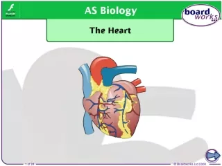
The human heart. The heart is a muscular organ located between the lungs in the centre of the chest (thorax), and is about the size of a fist. It pumps blood continuously around the body. An organism can lose consciousness within just a few seconds if the brain is deprived of blood.

The human heart
E N D
Presentation Transcript
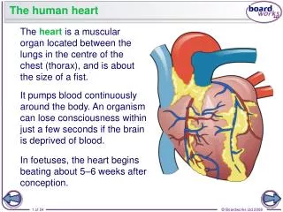
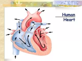
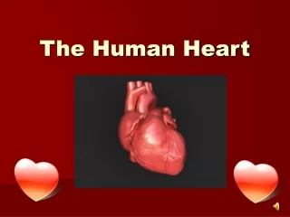
The human heart The heart is a muscular organ located between the lungs in the centre of the chest (thorax), and is about the size of a fist. It pumps blood continuously around the body. An organism can lose consciousness within just a few seconds if the brain is deprived of blood. In foetuses, the heart begins beating about 5–6 weeks after conception.
Cardiac muscle The heart mainly consists of cardiac muscle tissue, which like smooth muscle (but not skeletal muscle), contracts involuntarily. Cardiac muscle is made up of cells that are connected by cytoplasmic bridges. This enables electrical impulses to pass through the tissue. It contains large numbers of mitochondria and myoglobin molecules.
Preventing backflow Blood always flows in the same direction as it moves through the heart during each circulation of the body. Why is it important that blood does not flow backwards?
Heart valves Semilunar valve Semilunar valve Atrioventricular valve Atrioventricular valve The chambers of the heart are separated by valves which prevent blood from flowing in the wrong direction. There are valves between the atria and the ventricles… …and there are valves leading out of the ventricles.
How are valves held in place? The valves between the atria and ventricles are connected to the inner walls of the heart by tough tendons. valve open
How are valves held in place? The tendons allow the valves to close and hold the valve flaps in place. They prevent the valves from flipping up and turning inside out. Why is this important? valve closed valve open
Heart dissection – follow the instructions on the sheet. When you have finished, read about the cardiac cycle in the textbook and try to make notes about each of the stages on the worksheet from last lesson.
Cardiac output The amount of blood pumped around the body is called the cardiac output, and depends on two factors: • the stroke volume – the volume of blood pumped by the left ventricle in each heart beat. A typical value for an adult at rest is 75ml. • the heart rate – the number of times the heart beats per minute. A typical value for an adult at rest is 70bpm. cardiac output = stroke volume × heart rate A typical resting cardiac output is 4–6 litres per minute. This can rise to as much as 40 litres per minute in highly trained endurance athletes.
Activities • Card sort – put the statements into the correct order to show the events that occur during the cardiac cycle. • Try the questions on your worksheet. You may wish to use the textbook to help you interpret the information on the graph.
Pacemaker cells of the heart The heart can beat without any input from the nervous system as longs as its cells stay alive. This is due to myogenic contraction. Muscle cells (myocytes) in the heart have a slight electrical charge across their membrane. They are polarized. When the charge is reversed, they are said to be depolarized and this causes them to contract. Depolarization is initiated in a region of the heart called the sinoatrial node (SAN) – also known as the pacemaker – which is in the wall of the right atrium.
Artificial pacemakers Artificial pacemakers are devices implanted in people whose heart’s electrical conduction system is not working properly. Problems include the SAN not firing, and the blockage or disruption of impulses between the SAN and AVN, or in the bundle of His. Pacemakers monitor the heart’s electrical activity and stimulate the ventricles or atria to contract when necessary. Impulses are transmitted down electrodes implanted in the muscular walls.
What are electrocardiograms? The electrical activity of the heart can be monitored by an electrocardiograph. Several electrodes are attached to specific places on a person’s chest and limbs. These detect changes in polarization in the heart by measuring current at the skin surface. The leads are connected to a machine that draws an electrocardiogram (ECG).
ECG in diagnosis ECGs are used to diagnose problems with the heart, as variations in different components of the trace can indicate a disease or other abnormality. An ECG may be taken while the patient is relaxed or it may be taken before, during and after exercise. This is called a ‘stress test’ and usually involves the patient exercising on a treadmill while attached to an ECG machine.