The Human Heart
170 likes | 410 Views
The Human Heart. Structures & Functions. *Note…. Words highlighted in blue or red indicate the oxygenation of the blood flow through that structure. RED =oxygenated blood BLUE =deoxygenated blood L side heart=oxygenated blood R side heart=deoxygenated blood. Heart.

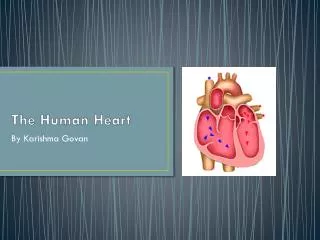
The Human Heart
E N D
Presentation Transcript
The Human Heart Structures & Functions
*Note… • Words highlighted in blue or red indicate the oxygenation of the blood flow through that structure. • RED=oxygenated blood • BLUE=deoxygenated blood • Lside heart=oxygenated blood • R side heart=deoxygenated blood
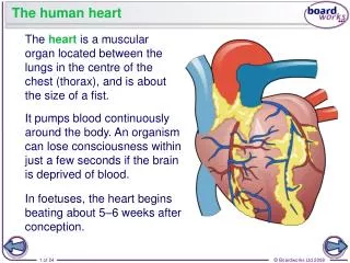
Heart Functions to pump blood to all parts of the body Muscle tissue = myocardium Size of a closed fist 5” long and 3.5” wide Weighs about 12-13 oz
Heart • Located in the Thoracic Cavity between the lungs, in front of the thoracic vertebrae and above the diaphram. • Lies centrally located, but the apex is slightly to the left midline. • Apex
Structures • Pericardium • Myocardium • Endocardium • Ventricular Septum • 4 chambers: R & Latria R & L ventricles
Heart • Structure- • Pericardium: double layer of fibrous tissue (outer layer) • Myocardium: cardiac muscle tissue (middle layer) • Endocardium: smooth muscle tissue lining the interior
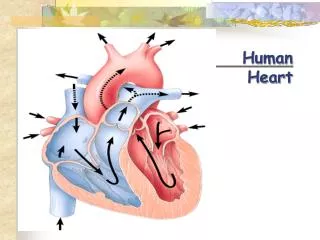
Structures leading to and from the heart • Superior & Inferior Vena Cava: large veins which brings deoxygenated blood to the R atrium from all parts of the body. • Pulmonary Artery: takes blood away from the R ventricle to the to the lungs for oxygen.
Structures leading to and from the heart…cont’ • Pulmonary veins: which bring oxygenated blood to the heart from the lungs. • Aorta: takes blood away from the L ventricle to the rest of the body.
Valves of the Heart • Tricuspid Valve: structure that is positioned between the R atrium and R ventricle. It has this name because there are 3 cusps (points) of attachment. It allows blood to flow from the R atrium into the R ventricle, but not in the opposite direction.
Valves of the Heart…cont’ • Mitral Valve: located between the L atrium and L ventricle. Blood flows from the L atrium to the L ventricle, while backflow from the ventricle to the atrium is prevented. • Closing of the valves produces heart sounds “lubb-dubb”
Valves of the Heart…cont’ • Semilunar Valves • Aortic Semilunar Valve: is at the orafice of the aorta. This valve permits blood flow out of the L ventricle to the aorta, but not backwards into the L ventricle. • Pulmonary Semilunar Valve: is found at the orafice of the pulmonary artery. It lets blood flow from the R ventticle into the pulmonary artery, and then into the lungs.
Labeled Structures Aorta Superior Vena Cava Pulmonary Artery R Atrium L Atrium Inferior Vena Cava I L Ventricle R Ventricle Ventricular Septum
Valves of the Heart Mitral Valve Tricuspid Valve Aortic Semilunar Valve Pulmonary Semilunar Valve
R & L Atria R & L Ventricle Pulmonary Artery Aorta Mitral Valve Tricuspid Valve Superior & Inferior Vena Cava Pulmonary Semilunar Valve Aortic Semilunar Valve Ventricular Septum *R side heart = deoxygenated blood *L side heart = oxygenated blood Summary