Download
1 / 47
470 likes | 497 Views
Explore the intricate processes of animal nutrition, covering topics like digestion, absorption of nutrients and water, energy, growth, and elimination. Learn about heterotrophs, intracellular and extracellular digestion, and the anatomy of digestive organs such as the stomach, small intestine, and large intestine. Discover how essential nutrients are absorbed and utilized in the body for energy and growth. Gain insights into common abnormalities and disorders related to the digestive system. Uncover the fascinating world of animal nutrition and its impact on overall health and well-being.

E N D
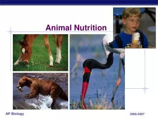
Animal nutrition Chapter 41
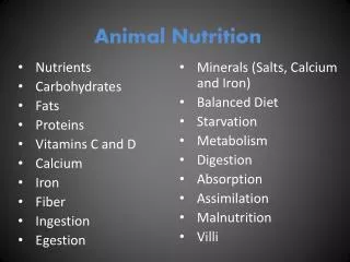
Function • 1. Digestion • 2. Absorption of nutrients/water • Energy • Growth • 3. Elimination
Fig. 41-9 Crop Gizzard Intestine Esophagus Pharynx Anus Mouth Typhlosole Lumen of intestine (a) Earthworm Foregut Midgut Hindgut Esophagus Rectum Anus Crop Mouth Gastric cecae (b) Grasshopper Stomach Gizzard Intestine Mouth Esophagus Crop Anus (c) Bird
General structure • Gastrointestinal tract (tube) • Mucosa: inner layer (epithelial) • Submucosa: connective • Muscularis: 2 layers of muscle • Serosa: outer layer (connective) • Plexues: nerves located in the submucosa
Digestive organs • Mouth • Pharynx • Esophagus • Stomach • Small intestine (duodenum, jejunum, ileum) • Large intestine (cecum, ascending colon, transverse colon, descending colon, sigmoid, rectum, anus)
Digestive organs • Accessory organs • Liver • Gallbladder • Pancreas
Digestion • Mouth • Teeth • Gizzard (in birds to help grind food) • Salivary glands • Secrete saliva • Amylase (enzyme to breakdown starch)
Digestion • Chew or mastication • Tongue pushes food • Pharynx • Epiglottis closes • Esophagus
Esophagus • Esophagus • Muscular tube • Connects pharynx to stomach • Peristalsis: • Rhythmic movement of muscle contractions • Moves food along • Esophageal sphincter: • End of esophagus keeps food in stomach
Stomach • Mucosa lining (epithelial) • Parietal cells • Secrete H + Cl ions • Chief cells • Secrete pepsinogen • Pepsinogen is converted to pepsin • Digests proteins
Stomach • Gastric juices: • HCl, pepsinogen & mucus pH=2 • Chyme: • Mixture of partially digested food
Small intestine • Chyme • Leaves stomach via pyloric sphincter • Duodenum • Digestive enzymes from pancreas • Bile from liver & gallbladder • Most digestion occurs in the duodenum & jejunum
Small intestines • Villi along intestine epithelium • Microvilli • “brush border” • Aids in absorption • Secretes enzymes • Break disaccharides (sucrose, lactose)
Accessory organs • Pancreas • Secretes fluids via pancreatic duct • Exocrine system • Trypsin & chymotrypsin (proteases) • Amylase (starch) • Lipase (fats) • Bicarbonate (neutralizes HCl) • Endocrine (insulin and glucagon)
Accessory organs • Liver • Secretes bile • Contains bile pigments & bile salts • Bile pigments are waste from break down of RBC • Eliminated
Accessory organs • Bile salts • Emulsify the fats • Bile made in liver • Stored in gall bladder • Released when eat fatty meal • Gallstones can block release
Smallintestines • Monosaccharides, aa are transported to the blood capillaries • Hepatic portal vein • Liver • Heart • Transported to body
Smallintestines • Fatty acids & monoglycerides • Villi • Triglycerides • Chylomicrons: (triglyceride & protein coat) • Lymph system
Fig. 41-15b Microvilli (brushborder) at apical(lumenal) surface Lumen Bloodcapillaries Epithelialcells Basal surface Epithelial cells Lacteal Lymphvessel Villi Key Nutrientabsorption
Fig. 41-16 Triglycerides Lumenof small intestine Fatty acids Monoglycerides Epithelialcell Triglycerides Phospholipids,cholesterol,and proteins Chylomicron Lacteal
Carbohydrate digestion Protein digestion Nucleic acid digestion Fat digestion Oral cavity,pharynx,esophagus Disaccharides Polysaccharides (starch, glycogen) (sucrose, lactose) Salivary amylase Smaller polysaccharides,maltose Stomach Proteins Pepsin Small polypeptides Essential nutrients Lumen ofsmall intes-tine DNA, RNA Fat globules Polypeptides Polysaccharides Pancreatic amylases Pancreatic trypsin andchymotrypsin Pancreatic nucleases Bile salts Maltose and otherdisaccharides Fat droplets Nucleotides Smallerpolypeptides Pancreatic lipase Pancreatic carboxypeptidase Glycerol, fattyacids, monoglycerides Amino acids Epitheliumof smallintestine(brushborder) Small peptides Nucleotidases Nucleosides Disaccharidases Dipeptidases, carboxypeptidase,and aminopeptidase Nucleosidasesandphosphatases Nitrogenous bases,sugars, phosphates Monosaccharides Amino acids
Large intestines • Absorbs water • Absorbs vitamin K • Concentrates wastes • E. coli • Feces • Cloaca • Combines feces & urine wastes in some animals
Food • BMR • Basal metabolic rate • Obesity • Heart disease, diabetes, stroke • Anorexia, Bulimia
Essential nutrients • Essential aa • Minerals • Vitamins • A, B-complex, C, D, E, K • Scurvy, rickets, pernicious anemia, bleeding
Abnormalities • Ulcers • H. pylori • Bacteria • Treated with antibiotics • Reflux: • Gastric juices go backwards to esophagus