Download
1 / 72
730 likes | 754 Views
Explore the preoperative and postoperative management options for NFPA, including observation, surgery, radiation therapy, and medical therapy. Learn about the prevalence, classification, and clinical manifestations of silent pituitary adenomas. Understand the biochemical and visual assessments, surgical indications, and surveillance guidelines. This review evaluates the conservative management of NFPAs and details essential assessments for micro and macroadenomas. Stay informed about the latest strategies for diagnosing and treating nonfunctional pituitary adenomas.

E N D
NONFUNCTIONAL PITUITARY ADENOMA S.BAHRI
Agenda • Introduction • Preoperative management of NFPA: • Observation • Surgical resection • Postoperative follow-up of NFPA • Post operative management of NFPA: • Surgical resection • Radiation therapy • Medical therapy • Conclusion
NFPA • Prevalence of pituitary adenoma is 80–100 cases per 100,000of the population. • NFPA accounts for 15–30% of these. • Annual incidence is 1 new case per 100,000 of the population. • Gonadotroph adenomas account for 25% to 35% of pituitary adenomas overall and 43% to 64% of silent adenomas. • Null-cell adenomas are nearly as common.
Nonfunctioning, or silent, pituitary adenoma • Clinically silent: • Can be classified by immunocytochemistry or can be detected by biochemical testing but do not cause clinical signs or symptoms. • Totally silent: • Can be classified by immunocytochemistry but do not secrete a sufficient amount of their hormonal products. • Null cell : Those that do not stain for any hormone by immunocytochemistry and do not secrete any hormonal products.
Gonadotroph Adenomas • In men: • An elevated FSH associated with low testosterone and LH that is not elevated. • An elevated serum FSH (with or without elevated alpha subunit) in up to one-quarter of these patients an isolated elevated alpha subunit concentration occurred in 7%. • An elevated testosterone and LH, with or without an elevated FSH • An increase in LH beta subunit in response to synthetic TRH
Gonadotroph Adenomas • In Pre menopause women: • Clinical manifestations : oligomenorrhea or amenorrhea and large ovarian cysts. • Biochemical testing: elevated FSH, normal or low LH, and markedly elevated estradiol. • In Post menopause women: • Elevated FSH concentration in the setting of a low or low normal LH • LH beta subunit response to synthetic TRH also occurs in women • LH beta subunit response to synthetic TRH also occurs in women.
MICROADENOMA • Essential biological assessment : • systematic prolactin and IGF1 assay is recommended in case of micro adenoma. • Systematic explore for cortisolhypersecretion is not recommended because there are no reported cases of infra clinical Cushing’s syndrome associated with pituitary microadenoma.
MICROADENOMA • Biochemical assessment: • Systematic exploration for anterior pituitary insufficiency is not recommended. • Exploration for anterior pituitary insufficiency, and for somatotropic insufficiency in particular, may be considered in6–9 mm pituitary micro incidentaloma if GH replacement therapy is likely to be prescribed.
MICROADENOMA • Visual assessment : • Not useful in suspected micro adenoma; • Nor is it recommended if the lesion is remote from the optic chiasm; • It is recommended if the lesion is close to the optic chiasm or is seen to be compressing it on MRI
SURGERY IN MICROADENOMA • Only progression indicates surgery; • No threshold size increase can be specified, • Age ,co morbidity and proximity to the chiasm are also to be taken into account.
MACROADENOMA • Biochemical assessment: • Screening for silent pituitary adenoma:
MACROADENOMA • Biochemical assessment: • Exploration of preoperative pituitary deficiency: • deficiency in at least one axis in 60–85% of cases. • Only the corticotroph axis should undergo dynamic exploration • The thyrotroph axis should undergo static exploration, TSH and free T4 assay. • In case of amenorrhea, even in women of menopausal age, FSH assay can guide diagnosis toward a peripheral or gonadotropin deficiency. • In male, LH, FSH and plasma total testosterone should be assayed. • Screening for somatotroph deficiency is not recommended
SURVEILLANCE IN MACROADENOMA • If there is a safety margin with respect to the chiasm (2–3 mm), especially in elderly patients and/or in case of co morbidity. • Invasion of cavernous space is not a reason for active primary treatment, as surgery will not cure the patient.
Objective: To characterize the natural history of patients with NFPAs managed conservatively. • Study Design : Retrospective analysis of patients presenting to a tertiary referral center between 1986 and 2009in London. • Study population : 66 patients were managed conservatively for a mean follow-up period of 4.3 years (range: 1–14.7). 47 (71%) had a macroadenoma, and 19 (29%) had a microadenoma.
Inclusion Criteria: • Patients with pituitary adenomas and no clinical or biochemical evidence of hormonal hypersecretion were included. • Exclusion Criteria: • Those presenting with apoplexy or a radiological follow-up period of less than 1 year were excluded.
Conclusion • Patients with NFPAs without optic chiasm compression can be managed conservatively. • All patients need pituitary function assessment, irrespective of tumour size.
Objective :To elucidate the natural history of nonfunctioning pituitary adenomas managed conservatively. • Study Design : Retrospective study between 2003 and 2013 in Queensland Australia . • Study population:50Patientswith nonfunctioning pituitary adenomas(mean age 49, range 17–85 years) who underwent at least 2 serial pituitary MRI scans over ≥6 months prior to any intervention
Primary end-point: was a ≥20% increase in volume or surgery. • Secondary end-points: were rate of pituitary dysfunction and pituitary apoplexy. • Exclusion Criteria: • (i) age younger than 16 years at baseline; • (ii) functioning adenoma, • (iii) radiological appearance suggestive of pituitary pathology other than pituitary adenoma, • (iv) less than6 months of serial imaging; • (v) previous surgical resection or radiation therapy • (vi) presentation with mass effect, such as visual field defect or pituitary apoplexy.
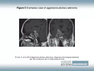
RESULTS • A high proportion of patients with NF macro adenomas without visual deficit at presentation eventually demonstrate an increase in volume and require surgical intervention. • In contrast, micro adenomas rarely progress; fewer than 10% increase in volume by ≥20% after 3–5 years of follow-up in this study.
RESULTS • A growth rate of >10 mm3/month over the initial 2 years of follow-up was highly predictive of either requiring surgery or ultimately exceeding a 20% increase in tumour volume. • Larger tumours at baseline were more likely to grow.
Conclusion • In macro adenomas where an increase in volume of more than 10 mm3/month occurs across the first 2 years of follow-up, consideration should be given to surgical intervention before visual or hormonal deficits arise. • Conversely, micro adenomas may be safely imaged at less frequent intervals • Based on our data, a micro adenoma which has not changed at 2 years may not need further imaging.
SURGERY IN MACROADENOMA Asymptomatic macro adenoma • Tumor progression (no specified threshold) • Non-compliant patient liable to escape follow-up; • Associated NFPA, male gender , proximity to chiasm and anticoagulation therapy, as this entails elevated risk of apoplexy • Patient age: In young patients without awaiting progression, Given the low risk of postoperative visual impairment; Risk of postoperative recurrence was lower with early surgery
SURGERY IN MACROADENOMA • Symptomatic adenoma • Visual disorders : • Improved vision is reported in some 80–90% of cases (including both partial and total recovery) • Recovery may be progressive, over a period of up to 1 year after surgery. • Some studies highlighted a correlation between percentage recovery and duration and severity of campimetric disorder
SURGERY IN MACROADENOMA • Pituitary deficiency : • The risk of onset of further pituitary deficiency associated with macro adenoma is estimated at 12% per year. • Surgery provided recovery of normal anterior pituitary function in about 30% of cases, at a mean of 1-year follow-up. the rate was higher for earlier management. • The risk of postoperative deterioration in pituitary function is about 10%.
SURGERY IN MACROADENOMA • Headaches : • classically due to distension of the dural envelopes • Retro-orbital or vertex locations suggest pituitary etiology.
Young women with or without pituitary deficiency: • NFA is rare in young women • In a patient hoping for pregnancy, the decision to operate may be based on gonadotroph deficiency • surgery provides pituitary recovery in about 30% of cases, while deficiency is found at diagnosis in more than 80% of cases .
pregnancy • maximum pituitary gland height can increase by 12 mm during pregnancy • When the adenoma is close to the optic pathways surgery should be considered; • In abstention from surgery clinical surveillance should be at least 3 monthly, with visual field test and MRI without contrast
Factors for recurrence • Clinical factors: • The best identified recurrence factor is postoperative adenomatous residue. • Young age, gender and initial tumor size are non-systematic recurrence factors . • Histologic factors : • Prognosis is classically poorer in silent corticotroph adenoma . when it occurs, is earlier and more aggressive . • Prognosis in silent mixed GH-PRL adenoma is poor.
Factors for recurrence • Histologic factors: • Ki67 :predicted recurrence with high specificity(89%) but poor sensitivity (54%), which suggests that new thresholds need to be identified in these adenomas. • New studies showed Ki67 is not predictive of recurrence risk but could be an effective index of progression risk in postoperative adenomatous residue. • Expression of subtype 2 and 5 somatostatin receptors is lower in recurrent adenoma.
Postoperative follow-up of NFPA • Biochemical assessment : • In the immediate (7–10 days) postoperative period, the focus should be on the assessment and correction of corticotroph deficiency and on screening for DI. An alternative may be to systematically perform dynamic exploration 4–6 weeks post-surgery. Corticotroph deficiency may resolve within 3 months of surgery so we can repeat corticotroph axis exploration at 3 months in case of initial deficiency
Postoperative follow-up of NFPA • Biochemical assessment : • Thyrotroph and gonadotroph axes should be assessed during the postoperative period. Although systematically performed 4–6 weeks after surgery. In case of initial deficiency, it is therefore recommended to repeat exploration at 3, 6 and 12 months.
Postoperative follow-up of NFPA • Biochemical assessment : • In the absence of residual tumor and postoperative pituitary deficiency : there is no need to explore for secondary onset • Incase of tumoral recurrence or residual tumor progression : pituitary exploration should be repeated at least once yearly in the absence of complementary treatment • In case of complementary radiation therapy pituitary function test should be done, at least once yearly for at least 10 years
Postoperative follow-up of NFPA • Ophthalmologic follow-up : • In case of preoperative ophthalmologic abnormality: a check-up is made 3 months after surgery. The examination is repeated every 6 months until maximum improvement is achieved. • In the absence of any visual impairment on the first postoperative examination: follow-up can be stopped if there is no supra sellar residue close to the optic pathways. • If (RT) is performed:Systematic 6-monthly ophthalmologic monitoring during the first 4 years and then annually for 10 years should be implemented.
What treatment can be offered for residue or recurrence? • Surgical revision : • Between 30 and 48% of NFPAs undergo surgical revision on a TSS approach either early for large residue , or later during tumoral progression. • Surgery indication: • progressive residue accessible for complete resection. • In case of persistent or recurrent symptomatic optic pathway compression; to obtain anatomic conditions allowing stereotactic RT (3–5 mm)safety margins • In case of post-RT tumor progression.
What treatment can be offered for residue or recurrence? • Radiation therapy: • After postoperative RT, 10-year progression-free survival being greater than 90% in most series . • Indication for RT: • Aggressive tumors • Large supra or extra sellar residual tumor • Confirmed recurrence.