Download
1 / 27
330 likes | 570 Views
Learn the intricate process of cerebral and cerebellar development, from neural tube formation to brain vesicle differentiation and cortical growth patterns. Understand key structures and changes in the brain's evolution.

E N D
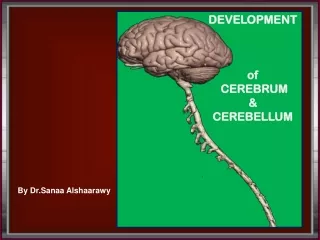
DEVELOPMENT of CEREBRUM &CEREBELLUM By Dr.Sanaa Alshaarawy
OBJECTIVES • By the end of the lecture the student should be able to: • Describe the formation of the neural tube. • List the 3 brain vesicles and their derivatives. • Describe the brain flexures. • Describe briefly the development of the cerebrum. • Describe briefly the development of the cerebellum.
INTRODUCTION By the beginning of the 3rd week of development, three germ cell layers become established, Ectoderm, Mesoderm and Endoderm.
EARLY DEVELOPMENT • During the middle of the 3rd week, the dorsal midline ectoderm undergoes thickening to form the neural plate (neuroectoderm). • The margins of the plate become elevated, forming neural folds. • So a longitudinal, midline depression, called the neural groove is formed. • The 2 neural folds then fuse together, thus sealing the neural groove and creating the neural tube.
Neural Tube DevelopmentThree-vesicles stage (End of 4th Week) • Formation of the neural tube is completed by the middle of the fourth week. • By the end of the 4th week, • Its upper end dilates & shows 3 vesicles: • Prosencephalon, • Mesencephalon, & • Rhombencephalon.
By the 5th weekfurther differentiation distinguishes five 2ry brain vesicles: • The prosencephalon divides into the two telencephalonand one diencephalonand • The Rhombencephalon divides into metencephalon andmyelencephalon.
Neural Tube DevelopmentFive-vesicles stage (5th week) Telencephalon Diencephalon Mesencephalon Metencephalon Myelencephalon
By the 4th week: • The neural tube grows rapidly and bends ventrally, producing two flexures: • Midbrain flexure: between the prosencephalon & the mesencephalon (midbrain) • Cervical flexure: • Between the hind brain & the spinal cord. Brain Flexures • Later Pontine flexureappears in the hindbrain, in the opposite direction, resulting in thinning of the roof of the hindbrain. Mesencephalon Rhobencephalon(hindbrain) Prosencephalon
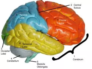
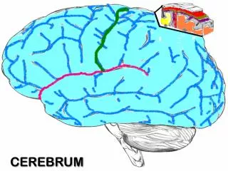
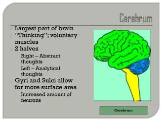
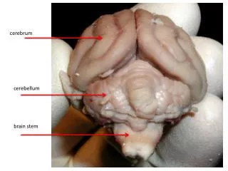
Development of the Cerebrum The cerebrum develops from the Telencephalon
Differentiation of Forebrain Vesicle • The (prosencephalon) or the forebrain vesicle differentiates into a: • Median part, ( diencephalon), • Two lateralcerebral vesicles or (telencephalic vesicles.) • The lumen gives the 2 lateral ventricles and the 3rd ventricle. • Both cavities communicating with each other through a wide interventricular foramen. • The cerebral hemispheres expand in all directions. • Its medial wallbecomes thin, flat and it is the site of choroid plexus of the lateral ventricle.
Development of the Cerebrum • The wall of the telencephalon is formed of 3 layers : • Ependymal (lining the cavity of the lateral ventricle). • Mantel;nerve cells forming the grey matter. • Marginal; nerve fibers forming the white matter.
As development proceeds the following changes occur: Most of the nerve cells migrate to the marginal layer forming the cerebral cortex. Some cells do not migrate and remains to form the basal ganglia.
Development of the Cerebrum • The cerebral hemispheres first appear on the day32 as a pair of bubble-like outgrowths of the Telencephalon. • By 16 weeks, the rapidly growing hemispheres are oval and have expanded back to cover the diencephalon.
By the end of the 3rd month the surfaces of the cerebral hemispheres are smooth. • By the 4th month the grey matter grows faster than the white matter, so, the cortex becomes folded into gyri separated by sulci. The gyri and sulci effectively increase the surface area of the brain. • The detailed pattern of gyri & sulci varies somewhat from individual to individual. 3rd month
Corpus striatum: • Itappears in 6th week in the floor of each cerebral hemisphere. • As the cerebral cortex differentiates and the fibers passing to and from it, pass through the corpus striatum, • The corpus striatum now divides into caudate nucleus & lentiform nucleus. • This fiber pathway forms the internal capsule.
Further expansion of cerebral hemisphere gives C-shape appearance to the hemisphere itself as well as its cavity (lateral ventricle). • Also the caudate nucleus elongates and assumes the shape of the lateral ventricle and remains related to it.
Development of the Cerebral Commissures • As the cerebral cortex develops, group of fibers, (commissures), connect the corresponding regions of the cortex. • These are: • Lamina terminalis. • Optic chiasma. • Anterior commissure. • Posterior commissure. • Hippocampal commissure. • Habenular commissure. • Corpus callosum.
Development of Insula The cortex covering the surface of the corpus striatum: grows relatively slowerthan the other cortices, so it is overgrown by the rest of the hemisphere and lies in the depth of the lateral sulcus. This is called the insula. So, the insular lobe is a portion of cerebral cortex that has invaginated to lie deep within the lateral sulcus.
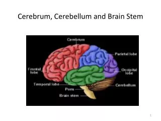
Development of the Cerebellum It develops from the dorsal part of the Metencephalon metencephalon myelencephalon
The metencephalondevelops into theponsand overlying cerebellum.
Development of the Cerebellum • Pontine flexure results in: • Moving the alar plates laterally then pending medially. • Stretching and thinning of the roof plate. • Widening of the cavity to form the 4thventricle. Alar plate
The dorsal parts thicken to form Rhombic lips, that will give rise to the cerebellum. Some neuroblasts migrate from the mantle layer to the marginal layer and form the cerebellarcortex. Others remains in the mantel layer and give rise to the cerebellar nuclei. The cerebellar peduncles develop lateras the axons of the neurones of the cerebellar nuclei grow out to reach the brain stem. Metencephalon: Changes in Alar plates Alar plate
As the cerebellar hemispheres develops they undergo a complicated process of transverse folding to form closely packed, leaf-like transverse gyri called folia. These processes of fissure formation and foliation continue throughout embryonic, fetal, and postnatal life, and they vastly increase the surface area of the cerebellar cortex. 35 d 50 d 90 d 150 d
Congenital Anomalies of The Brain • Mental retardation. • Seizures. • Cerebral palsy. • Cranium bifidum with or without meningocele & meningoencephalocele. • Microcephaly (abnormal smallness of the head, a congenital condition associated with incomplete brain development). • Agenesis of corpus callosum. • Hydrocephalus. • Arnold-Chiari malformation (herniated part of cerebellum through the foramen magnum leading to CSF obstruction ,so hydrocephalus results). • Anencephaly.
ANENCEPHALY In anencephaly, the brain and skull are minute and the infant does not usually survive.