Protein Structure and Function: Peptide Bond, Folding, Hierarchy
Explore the intricate details of protein structure, folding, and function, including peptide bond properties, favorable interactions, and secondary structure elements. Understand how proteins achieve their native fold and biological roles.

Protein Structure and Function: Peptide Bond, Folding, Hierarchy
E N D
Presentation Transcript
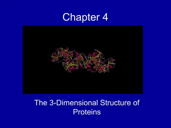
CHAPTER 4Proteins: Structure, Function, Folding • Structure and properties of the peptide bond • Structural hierarchy in proteins • Structure and function of fibrous proteins • Protein folding and denaturation • Structure analysis of globular proteins Key topics:
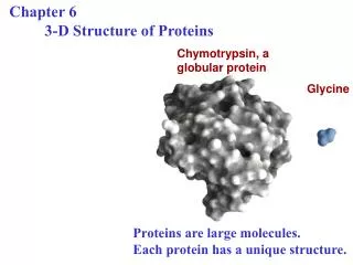
Structure of Proteins • Unlike most organic polymers, protein molecules adopt a specific 3-dimensional conformation in the aqueous solution. • This structure is able to fulfill a specific biological function • This structure is called the native fold • The native fold has a large number of favorable interactions within the protein • There is a cost in conformational entropy of folding the protein into one specific native fold
Protein Structures are compact Protein Sci. 2006 August; 15(8): 1829–1834. doi: 10.1110/ps.062305106.
Favorable Interactions in Proteins • Hydrophobic effect • Release of water molecules from the structured solvation layer around the molecule as protein folds increases the net entropy • Hydrogen bonds • Interaction of N-H and C=O of the peptide bond leads to local regular structures such as -helixes and -sheets • London dispersion • Medium-range weak attraction between all atoms contributes significantly to the stability in the interior of the protein • Electrostatic interactions • Long-range strong interactions between permanently charged groups • Salt-bridges, esp. buried in the hydrophobic environment strongly stabilize the protein
Structure of the Peptide Bond • Structure of the protein is partially dictated by the properties of the peptide bond • The peptide bond is a resonance hybrid of two canonical structures • The resonance causes the peptide bonds • be less reactive compared to e.g. esters • be quite rigid and nearly planar • exhibit large dipole moment in the favored trans configuration
The Rigid Peptide Plane and the Partially Free Rotations • Rotation around the peptide bond is not permitted • Rotation around bonds connected to the alpha carbon is permitted • f (phi): angle around the -carbon—amide nitrogen bond • y (psi): angle around the -carbon—carbonyl carbon bond • In a fully extended polypeptide, both y and f are 180°
Distribution of f and y Dihedral Angles • Some f and y combinations are very unfavorable because of steric crowding of backbone atoms with other atoms in the backbone or side-chains • Some f and y combinations are more favorable because of chance to form favorable H-bonding interactions along the backbone • Ramachandran plot shows the distribution of f and y dihedral angles that are found in a protein • shows the common secondary structure elements • reveals regions with unusual backbone structure 3
Secondary Structures • Secondary structure refers to a local spatial arrangement of the polypeptide chain • Two regular arrangements are common: • The helix • stabilized by hydrogen bonds between nearby residues • The sheet • stabilized by hydrogen bonds between adjacent segments that may not be nearby • Irregular arrangement of the polypeptide chain is called the random coil
The helix • The backbone is more compact with the y dihedral (N–C—C–N) in the range ( 0 < y < -70) • Helical backbone is held together by hydrogen bonds between the nearby backbone amides • Right-handed helix with 3.6 residues (5.4 Å) per turn • Peptide bonds are aligned roughly parallel with the helical axis • Side chains point out and are roughly perpendicular with the helical axis
The helix: Top View • The inner diameter of the helix (no side-chains) is about 4 – 5 Å • Too small for anything to fit “inside” • The outer diameter of the helix (with side chains) is 10 – 12 Å • Happens to fit well into the major groove of dsDNA • Residues 1 and 8 align nicely on top of each other • What kind of sequence gives an helix with one hydrophobic face?
Sequence Affects Helix Stability • Not all polypeptide sequences adopt -helical structures • Small hydrophobic residues such as Ala and Leu are strong helix formers • Pro acts as a helix breaker because the rotation around the N-Ca bond is impossible • Gly acts as a helix breaker because the tiny R-group supports other conformations
The Helix Macro-Dipole • Peptide bond has a strong dipole moment • Carbonyl O negative • Amide H positive • All peptide bonds in the helix have a similar orientation • The helix has a large macroscopic dipole moment • Negatively charged residues often occur near the positive end of the helix dipole
Sheets • The backbone is more extended with the y dihedral (N–C—C–N) in the range ( 90 < y < 180) • The planarity of the peptide bond and tetrahedral geometry of the -carbon create a pleated sheet-like structure • Sheet-like arrangement of backbone is held together by hydrogen bonds between the more distal backbone amides • Side chains protrude from the sheet alternating in up and down direction
Parallel and Antiparallel b Sheets • Parallel or antiparallel orientation of two chains within a sheet are possible • In parallelb sheets the H-bonded strands run in the same direction • In antiparallel b sheets the H-bonded strands run in opposite directions
Circular Dichroism (CD) Analysis • CD measures the molar absorption difference of left- and right- circularly polarized light: = L– R • Chromophores in the chiral environment produce characteristic signals • CD signals from peptide bonds depend on the chain conformation
b Turns (Hairpins) • b-turns occur frequently whenever strands in b sheets change the direction • The 180° turn is accomplished over four amino acids • The turn is stabilized by a hydrogen bond from a carbonyl oxygen to amide proton three residues down the sequence • Proline in position 2or glycine in position 3 are common in b-turns
Proline Isomers • Most peptide bonds not involving proline are in the trans configuration (>99.95%) • For peptide bonds involving proline, about 6-20% can be in the cis configuration • Proline isomerization is catalyzed by proline isomerases
Protein Tertiary Structure • Tertiary structure refers to the overall spatial arrangement of atoms in a polypeptide chain or in a protein • One can distinguish two major classes • fibrous proteins • ¤ typically insoluble; made from a single secondary structure • globular proteins • water-soluble globular proteins • lipid-soluble membraneous proteins
Fibrous Proteins: From Structure to Function Function Structure Example Tough, rigid, Cross-linked a-helixes a-keratin hard (nails, horns) Rigid linker (S—S) Tensile strength, Cross-linked triple-helixes Collagen non-stretching Flexible linker (Lys-HyLys) (tendons, cartilage) Soft, flexible Non-covalently held b-sheets non-stretchy van der Waals interaction Silk fibroin (egg sac, nest, web)
Structure of Collagen • Collagen is an important constituent of connective tissue: tendons, cartilage, bones, cornea of the eye • Each collagen chain is a long Gly- and Pro-rich left-handed helix • Three collagen chains intertwine into a right-handed superhelical triple helix • The triple helix has higher tensile strength than a steel wire of equal cross section • Many triple-helixes assemble into a collagen fibril
4-Hydroxyproline in Collagen • Forces the proline ring into a favorable pucker • Offer more hydrogen bonds between the three strands of collagen • The post-translational processing is catalyzed by prolyl hydroxylase and requires a-ketoglutarate, molecular oxygen, and ascorbate (vitamin C)
Silk Fibroin • Fibroin is the main protein in silk from moths and spiders • Antiparallel b sheet structure • Small side chains (Ala and Gly) allow the close packing of sheets • Structure is stabilized by • hydrogen bonding within sheets • London dispersion interactions between sheets
Spider Silk • Used for webs, egg sacks, and wrapping the prey • Extremely strong material • stronger than steel • can stretch a lot before breaking • A composite material • crystalline parts (fibroin-rich) • rubber-like stretchy parts
Motifs (folds) Arrangements of several secondary structure elements
Quaternary Structure Quaternary structure is formed by spontaneous assembly of individual polypeptides into a larger functional cluster
Structure of the Cro-DNA complex • 6Cro.pdb Albright, R. A. and B. W. Matthews (1998). "Crystal structure of lambda-Cro bound to a consensus operator at 3.0 A resolution." J Mol Biol 280(1): 137-51.
Protein Stability and Folding • A protein’s function depends on its three-dimensional structure. • Loss of structural integrity with accompanying loss of activity is called denaturation • Proteins can be denatured by • heat or cold; pH extremes; organic solvents • chaotropic agents: urea and guanidinium hydrochloride
Ribonuclease Refolding Experiment • Ribonuclease is a small protein that contains 8 cysteins linked via four disulfide bonds • Urea in the presence of 2-mercaptoethanol fully denatures ribonuclease • When urea and 2-mercaptoethanol are removed, the protein spontaneously refolds, and the correct disulfide bonds are reformed • The sequence alone determines the native conformation • Quite “simple” experiment, but so important it earned Chris Anfinsen the 1972 Chemistry Nobel Prize
How Can Proteins Fold So Fast? • Proteins fold to the lowest-energy fold in the microsecond to second time scales. How can they find the right fold so fast? • It is mathematically impossible for protein folding to occur by randomly trying every conformation until the lowest energy one is found (Levinthal’s paradox) • Search for the minimum is not random because the direction toward the native structure is thermodynamically most favorable
Protein Structure Methods: X-Ray Crystallography Steps needed: • Purify the protein • Crystallize the protein • Collect diffraction data • Calculate electron density • Fit residues into density Pros: • No size limits • Well-established Cons: • Difficult for membrane proteins • Cannot see hydrogens
Proton NMR spectrum of a protein Amides Aromatics Alphas Aliphatics Methyls
Structure Methods: Biomolecular NMR Steps needed: • Purify the protein • Dissolve the protein • Collect NMR data • Assign NMR signals • Calculate the structure Pros: • No need to crystallize the protein • Can see many hydrogens Cons: • Difficult for insoluble proteins • Works best with small proteins
Chapter 4: Summary In this chapter, we learned about: • the two most important secondary structures: • helixes • sheets • how properties and function of fibrous proteins are related • how to determine three-dimensional structures of proteins • one of the largest unsolved puzzles in modern biochemistry: how proteins fold?