Download

1 / 22
220 likes | 240 Views
Effects of Bisphenol A on Cancer Cell Proliferation. Anthony DiBello Central Catholic High School Grade 11. Cancer Overview. Cancer cells grow and divide at an unregulated pace (also known as proliferation ).

E N D
Effects of Bisphenol A on Cancer Cell Proliferation Anthony DiBello Central Catholic High School Grade 11
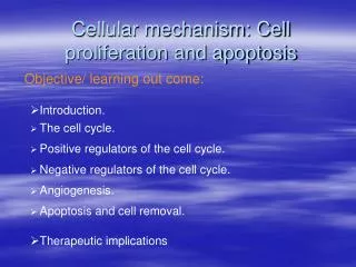
Cancer Overview • Cancer cells grow and divide at an unregulated pace (also known as proliferation). • Apoptosis (programmed cell death) does not occur in cancer cells. Extended passing on of this mutation eventually causes tumors to form. • Tumors can be metastatic (invasive of other body tissues) or benign(relatively harmless).
MG 63 Cell Line • Human cancer cell line • Osteosarcoma cells (aggressive form of bone cancer) • Useful model for testing the effects of variables on the proliferation of cancer cells
Bisphenol A • Originally produced as a synthetic estrogen • Endocrine-Disrupting Chemical used in the production of: • Polycarbonate plastic (beverage bottles, infant feeding bottles, food containers) • Epoxy resins (protective lining for canned food)
Bisphenol A and Cancer • As a xenoestrogen, BPA may contain chemical factors that either cause cancer, promote cancerous growth, inhibit cancer growth, or alter the progression/phenotype of cancer. • Some cancers are promoted by the work of xenohormones (including xenoestrogens). • Mimic the actions of a certain hormone (estrogen) • Might promote cancer growth
Purpose • To determine the effect of BPA exposure on cancer cell proliferation
Hypotheses • Null: BPA exposure WILL NOTsignificantly affect cancer cell proliferation • Alternative: BPA exposure WILLsignificantly affect cancer cell proliferation
Materials • 75 ml culture flask • Incubator • Nikon Inverted Microscope • Laminar Flow Hood • Laminar Flow Hood UV Sterilizing Lamp • Labeling Tape • Hemocytometer • Sterile PBS • Ethanol (70% and 100%) • Purple Nitrile gloves • Trypsin-EDTA • Pen/strep • Cryotank • Three 75 cm2 tissue culture treated flasks • Twelve 25 cm2tissue culture treated flasks • Fetal bovine serum (FBS) • MG63 Osteosarcoma Cancer Cell Line • Macropipette + sterile macropipette tips (1 mL, 5 mL, 10, mL, 20 mL) • Micropipettes + sterile tips • DMEM Serum - 1% and Complete Media (4 mM L-glutamine, 4500 mg/L glucose, 1 mM sodium pyruvate, and 1500 mg/L sodium bicarbonate + [ 10% fetal bovine serum for complete]) • 10 mg Bisphenol A
Procedure: Cell Culture • MG 63 cells were thawed and grown for four days. • When flasks reached a density of approximately 1 million cells/mL, the cells were passed into two flasks and incubated for 2 days at 37° C, 5% CO2.
Procedure: Proliferation Experiment – Day 0 (Addition of Variable) • Cells were trypsinized and suspended in 20 mL of media, creating a density of approximately 1x10^5 cells per flask • 0.5 ml of the cell suspension was added to 25 cm2 tissue culture treated flasks containing 4.5 mL of DMEM (com) media. • Two stock solutions of BPA were created using pure ethanol and DMEM media: 10^-4 M and 10^-5 M. • Experimental flasks were exposed to Bisphenol A and all flasks were incubated. (2 flasks per concentration x 3 concentrations x 2 days = 12 flasks total) • The cells were incubated at 37°C, 5% CO2 for the remainder of the study.
Procedure: Cell Density • Day 1 and Day 2 ▫ For each flask, cell densities were determined asfollows: • The cells were trypsinized and collected into cell suspension. • 10 μl aliquots were transferred to a Hemocytometer for quantification (eight counts per flask).
Images: Day 1 0 M Low (10^-6 M) High (10^-5 M)
Images: Day 2 0 M 10^-6 M 10^-5 M
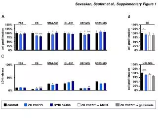
Conclusions • The effect of BPA exposure on cancer cell proliferation was significantfor both concentrations on day two only. • Null hypothesis rejected.
Future Changes • It is unlikely that all cell suspensions were perfectly homogenous. • Pipetting at various stages of experimentation was not perfectly synchronized. • BPA needed to be dissolved in pure ethanol, potentially harming cell growth. • Different exposures of BPA can be used (longer exposures and greater concentrations) • Test BPA exposure on other cell lines (C2C12, 3T3) • Use an Ames test to determine mutagenic capabilities of BPA on a non-cancer cell model. Limitations Extensions
References • Mark Krotec, PTEI • Phil Campbell, Ph.D. • Donald B. DeFranco, Ph.D. • https://www.ncbi.nlm.nih.gov/pmc/articles/PMC2967230/#!po=81.6667 • http://www.northcarolinahealthnews.org/2012/04/02/local-scientists-in-the-middle-of-the-bpa-debate/ • https://www.scientificamerican.com/article/just-how-harmful-are-bisphenol-a-plastics/