Download
1 / 5
50 likes | 89 Views
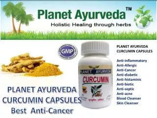
Cancer is a hyper proliferative disorder that metastasize into the vital organs in the body through invasion<br>followed by angiogenesis and distant metastasis. Curcumin suppresses the proliferation of a wide variety of tumor<br>cells, including breast carcinoma, colon carcinoma, renal cell carcinoma, hepatocellular carcinoma etc. The<br>poor bio availability is the main drawback of the regular curcumin, which was addressed by Aurea biolabs and<br>made a unique bio available formulation known as “cureitâ€. The cytotoxic effect of cureit was established by a<br>spectrophotometrical study using MTT on the effects of cureit on cell proliferation. It is inferred that the test sample<br>– “cureit†could serve as an anti cancer medication. <br>

E N D
Available online www.jocpr.com Journal of Chemical and Pharmaceutical Research, 2014, 6(9):96-100 ISSN : 0975-7384 CODEN(USA) : JCPRC5 Research Article Cell culture study on the cytotoxic effects of “Cureit”- a novel bio available curcumin-anti cancer effects Sreeraj Gopi*a, Robin Georgea, Shintu Judea and V. T. Sriraamb aR & D Centre, Aurea Biolabs (P) Ltd – A Plant Lipids Company, Cochin bAurous HealthCare Research and Development India Private Limited, Chennai _____________________________________________________________________________________________ ABSTRACT Cancer is a hyper proliferative disorder that metastasize into the vital organs in the body through invasion followed by angiogenesis and distant metastasis. Curcumin suppresses the proliferation of a wide variety of tumor cells, including breast carcinoma, colon carcinoma, renal cell carcinoma, hepatocellular carcinoma etc. The poor bio availability is the main drawback of the regular curcumin, which was addressed by Aurea biolabs and made a unique bio available formulation known as “cureit”. The cytotoxic effect of cureit was established by a spectrophotometrical study using MTT on the effects of cureit on cell proliferation. It is inferred that the test sample – “cureit” could serve as an anti cancer medication. Key words: Curcumin, tumor cells, cytotoxic effects, cancer _____________________________________________________________________________________________ INTRODUCTION Cancer is a hyperproliferative disorder that metastasize into the vital organs in the body through invasion followed by angiogenesis and distant metastasis. Within the last 25 years, much has been learned about the biochemical pathway that ultimately leads to cancer[1-3]. During the last decade, there has been extensive investigation of how Curcumin affects this overall process of tumorigenesis[4]. Figure1: Cancer in the body Anti Proliferative Effects of Curcumin Curcumin suppresses the proliferation of a wide variety of tumor cells, including breast carcinoma, colon carcinoma, renal cell carcinoma, hepatocellular carcinoma, T cell leukemia, B cell lymphoma, acute myelogenous leukemia, basal cell carcinoma, melanoma and prostate carcinoma. Additionally curcumin suppresses 96
Sreeraj Gopi et al ______________________________________________________________________________ J. Chem. Pharm. Res., 2014, 6(9):96-100 the proliferation of certain normal cells such as hepatocytes, epithelial cells, human vascular endothelial cells (HVEC), human vascular smooth muscle cells (HVSMC), osteoclasts, peripheral bood mononuclear cells (PBMC) and T lymphocytes. Curcumin also inhibits the cell proliferation induced by growth factors[5-7]. Curcumin inhibits farnesyl protein transferase (FPTase): Ras proteins must be isoprenylated at a conserved cysteine residue near the carboxyl terminus (Cys- 186 in mammalian Ras p21 proteins) in order to extend their biological activity. Previous studies indicate an intermediate in the mevalonate pathway, most likely farnesyl pyrophosphate, is the donor of this isoprenyl group, and that using inhibitors of the mevalonate pathway could block the transforming properties of ras oncogene. Chen et al. examined the effects of curcumin on farnesyl protein transferase (FPTase). They found that partially purified farnesyl protein transferase (FPTase) capable of catalyzing the farnesylation of unprocessed Rasp21 proteins in vitro was inhibited by curcumin and its derivatives [6]. This is another potential mechanism by which curcumin could suppress cellular growth. Suppression of NF-κB activation by Curcumin Members of the NF-κB transcription factor family play a central role in various responses leading to host defense, activating a rapid progression of gene expression. These transcription factors are dimeric complexes composed of different members of the Rel/NF-κB family of polypeptides. This family is distinguished by the presence of a Rel homology domain of about 300 amino acids that displays a 35% to 61 % identity between various family members[8-11]. Although NF-κB is a ubiquitous transcription factor, it plays its critical role in the cells of the immune system, where it controls the expression of various cytokines and the major histocompatibility complex genes. The inappropriate regulation of NF-κB and its dependent genes have been associated with various pathological conditions including toxic/septic shock, graft vs host reaction, acute inflammatory conditions, acute phase response, viral replication, radiation damage, atherosclerosis, and cancer. Figure 2: Molecular Targets of Curcumin Bio Availability of Curcumin It was known from the literatures that, the potential health benefits of curcumin are limited by its poor solubility, low absorption from the gut, and rapid metabolism. There are many bio available curcumin formulations available in the market, employing various additives to improve the bio availability. Aurea biolabs (A plantlipids company) developed a novel bio available curcumin formulation, completely in turmeric matrix, and its cytotoxic potential against cancerous cells was studied. MTT Cell Proliferation Assay Measurement of cell viability and proliferation forms the basis for numerous in vitro assays of a cell population’s response to external factors. The reduction of tetrazolium salts is now widely accepted as a reliable way to examine cell proliferation. The action of dehydrogenase enzymes generate the reducing equivalents such as NADH and NADPH in the metabolically active cells, which causes the reduction of the yellow tetrazolium MTT 3-(4, 5- dimethylthiazol-2-yl)-2, 5-diphenyltetrazolium bromide). The resulting intracellular purple formazan can be solubilized and quantified by means of spectrophotometer. The MTT Cell Proliferation Assay measures the cell proliferation rate and conversely, when metabolic events lead to apoptosis or necrosis, the reduction in cell viability. The number of assay steps has been minimized as much as possible to expedite sample processing. The MTT 97
Sreeraj Gopi et al ______________________________________________________________________________ J. Chem. Pharm. Res., 2014, 6(9):96-100 Reagent yields low background absorbance values in the absence of cells. For each cell type the linear relationship between cell number and signal produced is established, thus allowing an accurate quantification of changes in the rate of cell proliferation. The measurement of cell proliferation is based on the ability of the mitochondrial succinate-terazolium reductase system to convert 3-(4,5- dimethylthiazol-2-yl)-2,5- diphenyltetrazolium bromide (MTT) to a blue colored formazan. The test denotes the survival cells after toxic exposure. Figure 3: MTT Assay Reagents and Materials Used ? Test Compound – “cureit”- Bio available curcumin ? MCF-7 (Breast Cancer Cell Line) ? LnCAP (Prostrate Cancer Cell Line) ? HEK 293 T (Normal human embryonic kidney cells) ? Minimal Essential Medium (MEM) ? RPMI (Rosewell Park Memorial Institute) medium ? DMEM (Dulbecco's modified Eagle's medium) ? FBS (Fetal Bovine Serum) ? MTT ? DMSO (Dimethyl sulfoxide) Instruments Used •Spectrophotometer •Incubator Methodology MCF-7 (Breast Cancer Cell Line) was cultured in 5ml of Minimum Essential Medium (MEM) supplemented with fetal bovine serum(10%), L-glutamine(3%), penicillin (100IU/ml), streptomycin (100 µg/ml), amphotericin B (20 µg/ml), and phenol red. The pH of the medium was adjusted to 7.2-7.4 with 7.5% sodium bicarbonate and the flasks were incubated at 37°C in a humidified incubator (5% CO2/95% O2). LnCAP (Prostrate Cancer Cell Line) was cultured in RPMI (Roswell Park Memorial Institute) medium 1640 at 37°C in a humidified incubator (5% CO2/95% O2). HEK 293 (Normal human embryonic kidney cells) was cultured in DMEM (Dulbecco’s Modified Essential Medium) at 37°C in a humidified incubator (5% CO2/95% O2). The cytotoxic effect of the test compound was studied on this non-cancerous cell line also. All three cell cultures were treated with the test compound – “cureit”. 48 hours post treatment, the cells were treated with 100% MTT in media for 4 hours at 37°C in a humidified incubator (5% CO2/95% O2). The media was aspirated and the adherent cells 98
Sreeraj Gopi et al ______________________________________________________________________________ J. Chem. Pharm. Res., 2014, 6(9):96-100 were dissolved in DMSO. This was centrifuged at 5000RPM for 15 minutes to remove debris. Then the absorbance was measured spectrophotometrically at 570nm. RESULTS AND DISCUSSION The following is the result of the assay performed. Table 1: Cytotoxic Effect of Curcumin in MCF-7 cells Sample OD at 570nm Test sample (µg/ml) Batch I Batch II Control 0.5891 0.6512 10 0.6689 0.5912 20 0.4865 0.5012 40 0.3561 0.36941 80 0.2564 0.3251 150 0.2213 0.2512 Table 2: Cytotoxic Effect of Curcumin in LnCAP cells Sample OD at 570nm Batch I Batch II Batch III Control 0.7821 0.6891 0.5822 10 0.5676 0.6621 0.7012 20 0.5541 0.751 0.5421 40 0.4568 0.4987 0.4751 80 0.3211 0.3395 0.35005 150 0.4156 0.3564 0.32 Table 3: Cytotoxic Effect of Curcumin on HEK293 T cells Sample OD at 570nm Batch I Batch II Batch III Control 0.6453 0.5643 0.5666 10 0.7012 0.6972 0.6654 20 0.6754 0.7825 0.5671 40 0.6855 0.5769 0.5563 80 0.6321 0.5911 0.5612 150 0.3218 0.3092 0.5643 The effect of the test compound – Curcumin was studied on the growth kinetics of MCF-7, LnCAP and HEK 293 T cells using MTT Assay. Dose response for the test compound- curcumin was studied in the cell lines in concentrations ranging from 10-150 µg/ml. MCF-7 cells seem to be more susceptible to the test sample induced cytotoxicity compared to LnCAP cells. It is inferred that the test sample – “cureit” could serve as an anti cancer medication. CONCLUSION The anti cancer properties of curcumin are already known in the scientific world. The poor bio availability is the main drawback of the regular curcumin, which was addressed by Aurea biolabs and made a unique bio available formulation known as “cureit”. The cytotoxic effect of cureit was established and it is inferred that the test sample – “cureit” could serve as an anti cancer medication. REFERENCES [1]Dobelis Hamper IN (ed): Magicand Medicine of Plants.Pleasantville, NY, Reader’s Digest Association, 1986. [2]Srimal RC, Dhawan BN: J Pharm Pharmacol , 1973, 25(6), 447–452. [3]Jain SK, DeFilipps RA: Medicinal Plants of India. Algonac, MI, Reference, 1991, p 120 [4]Nadkarni AK: Indian Materia Medica, Vol 1,Bombay, India, Popular Book Depot, 1954 [5]Chang HM, But BPH: Pharmacology and Applications of Chinese Materia Medica, Vol 2, Philadelphia, PA, World Scientific, 1986, 936–939. [6]Tu G, Fang Q, Guo J, Yuan S, Chen C, Chen J, Chen Z, Cheng S, Jin R, Li M, et al.: Pharmacopoeia of the People’s Republic of China. Guangzhou, P.R. China, Guangdong Science and Technology Press, 1992, 202–203. Batch III 0.6222 0.6433 0.4621 0.3625 0.3625 0.2021 Avg 0.6208 0.6345 0.4833 0.3627 0.3147 0.2249 % Inhibition -2.20 22.15 41.58 49.31 63.78 %SD 6.24 4.09 1.8 17 11 Avg 0.6845 0.6436 0.6157 0.4769 0.3369 0.3640 % Inhibition -3.68 0.82 23.19 45.73 41.37 % SD 10.6 19.05 4.35 4.4 13.26 Avg 0.5921 0.6879 0.6750 0.6062 0.5948 0.3984 % Inhibition -10.81 -8.73 2.35 4.19 35.82 % SD 7.8 2.9 16 11.5 6.0 36.1 99
Sreeraj Gopi et al ______________________________________________________________________________ J. Chem. Pharm. Res., 2014, 6(9):96-100 [7]Leung A: Encyclopedia of Common Natural Ingredients Used in Food, Drugs, and Cosmetics,New York, Wiley, 1980, 313– 314. [8] Lampe V, Milobedeska J, Kostanecki V: Ber Dtsch Chem Ges , 1910, 43,21-63, [9]Lampe V, Milobedeska J: Ber Dtsch Chem Ges, 1913, 46, 22-35. [10]Ammon HP, Wahl MA: Planta Med 57(1):1–7, 1991 [11]Cheng AL, Hsu CH, Lin JK, Hsu MM, Ho YF, Shen TS, Ko JY,Lin JT, Lin BR, Ming-Shiang W, Yu HS, Jee SH, Chen GS, Chen TM, Chen CA, LaiMK, Pu YS, PanMH,Wang YJ, Tsai CC, Hsieh CY: Anticancer Res, 2001, 21(4B):2895–2900. 100