Download

1 / 63
640 likes | 718 Views
Understand the divisions of the nervous system, neuron types, properties, structures, and glial cells. Learn about neural transmission speed, myelin sheath disorders, and nerve fiber regeneration. Discover the roles of different types of neurons and the functions of sensory and motor divisions.

E N D
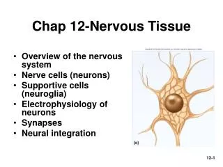
Chapter 12Nervous Tissue • Overview of the nervous system • Cells of the nervous system • Electrophysiology of neurons • Synapses • Neural integration
Subdivisions of the Nervous System Two major anatomical subdivisions • Central nervous system (CNS) • brain & spinal cord enclosed in bony coverings • Peripheral nervous system (PNS) • nerve = bundle of nerve fibers in connective tissue • ganglion = swelling of cell bodies in a nerve
Functional Divisions of PNS • Sensory (afferent) divisions (receptors to CNS) • visceral sensory division • somatic sensory division • Motor (efferent) division (CNS to effectors) • visceral motor division (Autonomic NS) effectors: cardiac, smooth muscle, glands • sympathetic division (action) • parasympathetic division (digestion) • somatic motor division effectors: skeletal muscle
Fundamental Types of Neurons • Sensory (afferent) neurons • receptors detect changes in body and external environment • this information is transmitted into brain or spinal cord • Interneurons (association neurons) • lie between sensory & motor pathways in CNS • 90% of our neurons are interneurons • process, store & retrieve information • Motor (efferent) neuron • send signals out tomuscles & gland cells • organs that carry out responses called effectors
Fundamental Properties of Neurons • Excitability (irritability) • ability to respond to changes in the body and external environment called stimuli • Conductivity • produce traveling electrical signals • Secretion • when electrical signal reaches end of nerve fiber, a chemical neurotransmitter is secreted
Structure of a Neuron • Cell body = soma • single, central nucleus with large nucleolus • cytoskeleton of microtubules & neurofibrils (bundles of actin filaments) • compartmentalizes RER into Nissl bodies • lipofuscin product of breakdown of worn-out organelles -- more with age • Vast number of short dendrites • for receiving signals • Singe axon (nerve fiber) arising from axon hillock for rapid conduction • axoplasm & axolemma & synaptic vesicles
Axonal Transport • Many proteins made in soma must be transported to axon & axon terminal • repair axolemma, for gated ion channel proteins, as enzymes or neurotransmitters • Fast anterograde axonal transport • either direction up to 400 mm/day for organelles, enzymes, vesicles & small molecules • Fast retrograde for recycled materials & pathogens • Slow axonal transport or axoplasmic flow • moves cytoskeletal & new axoplasm at 10 mm/day during repair & regeneration in damaged axons
Six Types of Neuroglial Cells • Oligodendrocytes form myelin sheaths in CNS • each wraps processes around many nerve fibers • Astrocytes • contribute to BBB & regulate composition of brain tissue fluid • most abundant glial cells - form framework of CNS • sclerosis – damaged neurons replace by hardened mass of astrocytes • Ependymal cells line cavities & produce CSF • Microglia (macrophages) formed from monocytes • concentrate in areas of infection, trauma or stroke • Schwann cells myelinate fibers of PNS • Satellite cells with uncertain function
Myelin Sheath • Insulating layer around a nerve fiber • oligodendrocytes in CNS & schwann cells in PNS • formed from wrappings of plasma membrane • 20% protein & 80 % lipid (looks white) • In PNS, hundreds of layers wrap axon • the outermost coil is schwann cell • Gaps between myelin segments = nodes of Ranvier • Initial segment (area before 1st schwann cell) & axon hillock form trigger zone where signals begin
Myelin Sheath • Note: Node of Ranvier between Schwann cells
Myelin Sheath Formation • Myelination begins during fetal development, but proceeds most rapidly in infancy. • Neurilemma: outermost coating of Schwann Cell
Diseases of the Myelin Sheath • Multiple Sclerosis (MS) Myelin sheath of CNS deteriorate and are replaced by scar tissue • Starts somewhere between 20s-40s, patients survive 7-32 years after the onset • Symptoms: depend on what part of CNS is involved: blindness, speech defects, tremors, neurosis • No cure, but it might be immune disorder triggered by a virus. Treatments are used to treat symptoms.
Speed of Nerve Signal • Speed of signal transmission along nerve fibers • depends on diameter of fiber & presence of myelin • large fibers have more surface area for signals • Speeds • small, unmyelinated fibers = 0.5 - 2.0 m/sec • small, myelinated fibers = 3 - 15.0 m/sec • large, myelinated fibers = up to 120 m/sec • Functions • slow signals supply the stomach & dilate pupil • fast signals supply skeletal muscles & transport sensory signals for vision & balance
Regeneration of Peripheral Nerve Fibers • Can occur if soma & neurilemmal tube is intact • Stranded end of axon & myelin sheath degenerate • Healthy axon stub puts out several sprouts • Tube guides lucky sprout back to its original destination
Electrical Potentials & Currents • Neuron doctrine -- nerve pathway is not a continuous “wire” but a series of separate cells • Neuronal communication is based on mechanisms for producing electrical potentials & currents • electrical potential - difference in concentration of charged particles between different parts of the cell • electrical current - flow of charged particles from one point to another within the cell • Living cells are polarized • resting membrane potential is -70 mV with a relatively negative charge on the inside of nerve cell membranes
Resting Membrane Potential • Unequal electrolytes distribution • diffusion of ions down their concentration gradients • selective permeability of plasma membrane • electrical attraction of cations and anions • Explanation for -70 mV resting potential • membrane very permeable to K+ • leaks out until electrical gradient created attracts it back in • membrane much less permeable to Na+ • Na+/K+ pumps out 3 Na+ for every 2 K+ it brings in • works continuously & requires great deal of ATP • necessitates glucose & oxygen be supplied to nerve tissue
Be clear on vocabulary • Polarize = to increase the difference in concentration. To move away from no electricity 0mV • Resting potential is polarized • There’s a difference in Na+/K+ conc. • Depolarize = To move toward no electricity • Allowing Na+/K+ to go where they want. • “Opening flood gates” • Repolarize = To go back to the original
Ionic Basis of Resting Membrane Potential • Na+ concentrated outside of cell (ECF) • K+ concentrated inside cell (ICF)
Local Potentials • Local disturbances in membrane potential • occur when neuron is stimulated by chemicals, light, heat or mechanical disturbance • depolarization decreases potential across cell membrane due to opening of gated Na+ channels • Na+ rushes in down concentration and electrical gradients • Na+ diffuses for short distance inside membrane producing a change in voltage called a local potential • Differences from action potential • are graded (vary in magnitude with stimulus strength) • are decremental (get weaker the farther they spread) • are reversible as K+ diffuses out, pumps restore balance • can be either excitatory or inhibitory (hyperpolarize)
Action Potentials • More dramatic change in membrane produced where high density of voltage-gated channels occur • trigger zone has 500 channels/m2 (normal is 75) • If threshold potential (-55mV) is reached voltage-gated Na+ channels open (Na+ enters causing depolarization) • Passes 0 mV & Na+ channels close (peaks at +35) • K+ gates fully open, K+ exits • no longer opposed by electrical gradient • until repolarization occurs • Negative overshoot produceshyperpolarization
Action Potentials • Called a spike • Characteristics of AP • follows an all-or-none law • voltage gates either open or don’t • nondecremental (do not get weaker with distance) • irreversible (once started goes to completion and can not be stopped)
The Refractory Period • Period of resistance to stimulation • Absolute refractory period • as long as Na+ gates are open • no stimulus will trigger AP • Relative refractory period • as long as K+ gates are open • only especially strong stimulus will trigger new AP • Refractory period is occurring only to a small patch of membrane at one time (quickly recovers)
Impulse Conduction in Unmyelinated Fibers • Threshold voltage in trigger zone begins impulse • Nerve signal (impulse) - a chain reaction of sequential opening of voltage-gated Na+ channels down entire length of axon • Nerve signal (nondecremental) travels at 2m/sec
Saltatory Conduction in Myelinated Fibers • Voltage-gated channels needed for APs • fewer than 25 per m2 in myelin-covered regions • up to 12,000 per m2 in nodes of Ranvier • Fast Na+ diffusion occurs between nodes
Saltatory Conduction of Myelinated Fiber • Notice how the action potentials jump from node of Ranvier to node of Ranvier.
Synapses Between Two Neurons • First neuron in path releases neurotransmitter onto second neuron that responds to it • 1st neuron is presynaptic neuron • 2nd neuron is postsynaptic neuron • Number of synapses on postsynaptic cell variable • 8000 on spinal motor neuron • 100,000 on neuron in cerebellum
The Discovery of Neurotransmitters • Histological observations revealed a 20 to 40 nm gap between neurons (synaptic cleft) • Otto Loewi (1873-1961) first to demonstrate function of neurotransmitters at chemical synapse • flooded exposed hearts of 2 frogs with saline • stimulated vagus nerve of one frog --- heart slows • removed saline from that frog & found it would slow heart of 2nd frog --- “vagus substance” discovered • later renamed acetylcholine
Chemical Synapse Structure • Presynaptic neurons have synaptic vesicles with neurotransmitter and postsynaptic have receptors
Postsynaptic Potentials • Excitatory postsynaptic potentials (EPSP) • a positive voltage change causing postsynaptic cell to be more likely to fire • result from Na+ flowing into the cell • glutamate & aspartate are excitatory neurotransmitters • Inhibitory postsynaptic potentials (IPSP) • a negative voltage change causing postsynaptic cell to be less likely to fire (hyperpolarize) • result of Cl- flowing into the cell or K+ leaving the cell • glycine & GABA are inhibitory neurotransmitters • ACh & norepinephrine vary depending on cell
Types of Neurotransmitters • 100 neurotransmitter types in 4 major categories • Acetylcholine • formed from acetic acid & choline • Amino acid neurotransmitters • Monoamines • synthesized by replacing -COOH in amino acids with another functional group • catecholamines (epi, NE & dopamine) • indolamines (serotonin & histamine) • Neuropeptides (next)
Neuropeptides • Chains of 2 to 40 amino acids • Stored in axon terminal as larger secretory granules Act at lower concentrations • Longer lasting effects • Some released from nonneural tissue • gut-brain peptides cause food cravings • Some function as hormones • modify actions of neurotransmitters
Monamines, • Catecholines: Come from amino acid tyrosine • Made in adrenal medulla • Blood soluable • Prepare body for activity • High levels in stressed people • Norepinephrine: raises heart rate, releases E • Dopamine: elevates mood • Helps with movement, balance • Low levels = Parkinson’s disease
Synaptic Transmission 3 kinds of synapses with different modes of action • Excitatory cholinergic synapse • Inhibitory GABA-ergic synapse • Excitatory adrenergic synapse Synaptic delay (.5 msec) • time from arrival of nerve signal at synapse to start of AP in postsynaptic cell
Other Catecholamines • Serotonin: sleepiness, alertness, thermoregulation, mood • Comes from tryptophan • Antidepressents: inhibit the reuptake, so it stays in the synaptic cleft longer. • Histamine: Sleep modulator • Vasodilation
Why Yawn? • caused by an excess of carbon dioxide & lack of oxygen in the blood? • Increase in the amount of catecholamines being released? • Herd Instinct so the group can synchronize sleep patterns?
Excitatory Cholinergic Synapse • Nerve signal opens voltage-gated calcium channels • Triggers release of ACh which crosses synapse • ACh receptors trigger opening of Na+ channels producing local potential (postsynaptic potential) • When reaches -55mV, triggers AP
Inhibitory GABA-ergic Synapse • Nerve signal triggers release of GABA (-aminobutyric acid) which crosses synapse • GABA receptors trigger opening of Cl- channels producing hyperpolarization • Postsynaptic neuron now less likely to reach threshold
Excitatory Adrenergic Synapse • Neurotransmitter is NE • Acts through 2nd messenger systems (cAMP) • Receptor is an integral membrane protein associated with a G protein, which activates adenylate cyclase, which converts ATP to cAMP • cAMP has multiple effects • synthesis of new enzymes • activating enzymes • opening ligand gates • produce a postsynaptic potential
Cessation & Modification of the Signal • Mechanisms to turn off stimulation • diffusion of neurotransmitter away from synapse into ECF where astrocytes return it to the neurons • synaptic knob reabsorbs amino acids and monoamines by endocytosis & breaks them down with monoamine oxidase • acetylcholinesterase degrades ACh in the synaptic cleft • choline reabsorbed & recycled • Neuromodulators modify synaptic transmission • raise or lower number of receptors • alter neurotransmitter release, synthesis or breakdown • nitric oxide stimulates neurotransmitter release
Neural Integration • More synapses a neuron has the greater its information-processing capability • cells in cerebral cortex with 40,000 synapses • cerebral cortex estimated to contain 100 trillion synapses • Chemical synapses are decision-making components of the nervous system • ability to process, store & recall information is due to neural integration • Neural integration is based on types of postsynaptic potentials produced by neurotransmitters