Download
1 / 95
950 likes | 1.02k Views
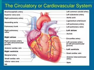
The Circulatory and Cardiovascular Systems Vocabulary Important info Headings. The Heart. Surface Projection of the Heart. Superior right point at the superior border of the 3 rd right costal cartilage

E N D
The Circulatory and Cardiovascular Systems Vocabulary Important info Headings
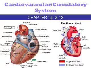
Surface Projection of the Heart • Superior right point at the superior border of the 3rd right costal cartilage • Superior left point at the inferior border of the 2nd left costal cartilage 3cm to the left of midline • Inferior left point at the 5th intercostal space, 9 cm from the midline • Inferior right point at superior border of the 6th right costal cartilage, 3 cm from the midline
Four chambers Atria Receiving chambers Right atrium Left atrium Ventricles Discharging chambers Right ventricle Left ventricle Heart valves Allow blood to flow in one direction ONLY Four valves Atrioventricular Valves – between atria and ventricles Bicuspid valve (left) Tricuspid valve (right) Semilunar valves between ventricle and artery Pulmonary semilunar valve Aortic semilunar valve The Heart: Internal Anatomy
Layers of Heart Wall • Epicardium • visceral layer of serous pericardium • Myocardium • cardiac muscle layer is the bulk of the heart • Endocardium • chamber lining & valves
Valve Function • Ventricles contract, blood pumped into aorta and pulmonary trunk through SL valves • Atria contract, blood fills ventricles through A-V valves
Heart Murmur • Heart murmurs are most often caused by defective heart valves. • A valve may be unable to close completely. • This leads to regurgitation, which is blood leaking backward through the valve when it should be closed Normal heartbeat murmur
Heart Sounds Where to listen on chest wall for heart sounds.
What Causes the Heartbeat? LUB DUP
Autorhythmic Cells Cells fire spontaneously, act as pacemaker and form conduction system for the heart SA node cluster of cells in wall of Rt. Atria begins heart activity that spreads to both atria excitation spreads to AV node AV node in atrial septum, transmits signal to Bundle of His AV bundle of His the connection between atria & ventricles divides into bundle branches & purkinje fibers, large diameter fibers that conduct signals quickly Conduction System of Heart
Rhythm of Conduction System • SA node fires spontaneously 90-100 times per minute • AV node fires at 40-50 times per minute • If both nodes are suppressed fibers in ventricles by themselves fire only 20-40 times per minute • Artificial pacemaker needed if pace is too slow • Extra beats forming at other sites are called Ectopic Pacemakers • caffeine & nicotine increase activity
Timing of Atrial & Ventricular Excitation • SA node setting pace since is the fastest • In 50 msec excitation spreads through both atria and down to AV node • 100 msec delay at AV node due to smaller diameter fibers- allows atria to fully contract filling ventricles before ventricles contract • In 50 msec excitation spreads through both ventricles simultaneously
Depolarization & Repolarization • Depolarization • Cardiac cell resting membrane potential is -90mv • excitation spreads through gap junctions • fast Na+ channels open for rapid depolarization • Plateau phase • 250 msec (only 1msec in neuron) • slow Ca+2 channels open, let Ca +2enter from outside cell and from storage in sarcoplasmic reticulum, while K+ channels close • Ca +2 binds to troponin to allow for actin-myosin cross-bridge formation & tension development • Repolarization • Ca+2 channels close and K+channels open & -90mv is restored as potassium leaves the cell • Refractory period • very long so heart can fill
Physiology of Contraction • Depolarization, plateau, repolarization
Electrocardiogram---ECG or EKG • EKG • Action potentials of all active cells can be detected and recorded • P wave • atrial depolarization • P to Q interval • conduction time from atrial to ventricular excitation • QRS complex • ventricular depolarization • T wave • ventricular repolarization
One Cardiac Cycle • At 75 beats/min, one cycle requires 0.8 sec. • Systole (contraction) and Diastole (relaxation) of both atria, plus the systole and diastole of both ventricles • End Diastolic Volume (EDV) • volume in ventricle at end of diastole, about 130 ml • End Systolic Volume (ESV) • volume in ventricle at end of systole, about 60 ml • Stroke Volume (SV) • the volume ejected per beat from each ventricle, about 70 ml • SV = EDV - ESV
Blood Pressure • Measurements by health professionals are made on the pressure in large arteries • Systolic – pressure at the peak of ventricular contraction (CONTRACTION OF HEART) • Diastolic– pressure when ventricles relax (RELAXATION OF HEART) • Pressure in blood vessels decreases as the distance away from the heart increases
Pulse • Pulse • Pressure wave of blood • “Pressure Points” • Area where pulse is easily palpated • Simple monitoring Figure 11.16
Variations in Blood Pressure • Human normal range is variable • Normal BP • 140–110 mm hg systolic • 80–75 mm hg diastolic • Hypotension • Low systolic (below 110 mm hg) • Often associated with illness • Hypertension • High systolic (above 140 mm hg) • Can be dangerous if it is chronic
Measuring Arterial Blood Pressure Figure 11.18
Nervous System: Big Brother • Nervous system controls heartbeat • Sympathetic NS= fight or flight • Parasympathetic NS = relaxation
What is your Resting Heart Rate? Normal = 60-75 Beats/Minute
The 3 Main Functions of Blood: • Transportation • Protection • Regulation • Blood is a connective tissue in liquid form • Greatest benefit from homeostasis: • Continuous flow of blood thru 60,000 miles of blood vessels
TRANSPORTATION: • Blood moves thru body where cells receive: • Nutrients from digestive organs • Oxygen from lungs • Hormones secreted from endocrine gland • Cells give blood waste • (CO2, urea & uric acid) & their secretions
Protection: • From harmful microorganism & their toxins • Through Phagocytic white blood cells • Specialized proteins called Antibodies • Against fluid loss after an injury by clotting
Figure This figure highlights some of the major acute (short-term) effects on the body during exercise. Regulation: • Regulates acid-base balance of the body fluids • By way of buffers • Neutralize potential harmful effects of: • too much co2 • actic acid • other compounds • Body temp. by cooling or heating parts of body • Controlled by Hypothalamus • Controls volume of blood flow to diff. areas of body
Properties of Blood: • Color • Volume • pH
COLOR • RED COLOR = • HEMOGLOBIN (PIGMENT PROTEIN) • Arterial blood the O2 molecules are chemically bound to hemoglobin • Crimson-red color • Venous blood O2 mol. are not as prevalent & blood= • Dark red color w/a slightly bluish tint • SEEN THROUGH SKIN VEINS LOOK GREENISH- BLUE but it is NOT GREEN OR BLUE
VOLUME • 8% OF BODY WEIGHT • Most in vessels--rest in heart • Does not vary much from day to day or year to year • Avg. Male = • 5-6 liters of blood • Avg. Female = • 4-5 liters of blood • Difference due to avg body weight not sex Apx. 8 pints
Blood is thicker, denser, & more adhesive than H2O • Due to formed elements (red blood cells) • Causes blood to flow 5x slower than H2O • Resistance to flow = viscosity • Blood is a viscous substance b/c it resists flow more than water Figure The shear rate dependence of normal human blood viscoelasticity at 2 Hz and 22 °C.
Slightly alkaline (aka: basic) • pH = 7.35-7.45 • Range stays small despite change in: • Diet • Cell secretions • Metabolic rateby buffering systems that remove h+ ions • If buffers fail: • BLOOD TOO ACIDIC (pH below 6.0) • Body cells stop functioning • No homeostasis = Acidosis • Too little acid in blood = Alkalosis (a lot less common)
Leukocytes • Less than 1% of total blood volume • 5000 TO 10,000 in cubic mm • Any change in number… • High or low indicates a disease
Types: • All contain a nucleus • (unlike the RBC’s) • Can wander outside the Circ. System • Wbc cells differ in: • Nature of cytoplasm • Size • Shape of nucleus • Response to different staining techniques • Divided into 2 groups by cytoplasm differences: • Granulocytes • Agranulocytes
Granulocytes • Cytoplasm contains highly visible pebble-like objects, known as granules • Twice the size of RBC’s • They contain a nucleus that is split into sections called lobes • Produced in red marrow
Three types: • Eosinophils • Neutrophils • Basophils • Names come from the type of stain that brings out their distinguishing features • Neutral • Eosin • Basic
Neutrophil: • Most abundant = granulocyte • Stain pink in a neutral stain • Nucleus contains: 2 to 5 lobes • Interconnected by thin bridges • Make up about 60% of all wbc’s in a normal blood sample
Eosinophils: • 1 to 4% of WBC’s in a normal blood sample • Granules stain red in an acid stain that contains a dye known as eosin • Nucleus = 2 lobes
Eosinophils are not: • Very mobile • Or active • But can phagocytize certain foreign particles produced by allergic reactions • Invading parasites • Pollen grains • Mold spores
Basophils: • Rarest0.5% or less of wbc’s in blood • Large granules that stain blue in basic stain • Nucleus is often bent into an s-shape with 2 lobes
Basophils & Mast cells produce a substance called = histamine • causes swelling or inflammation • Swelling tells other wbc’s where to find the site of infection ***Mast cells reside in tissues in the body, and basophils are in the blood stream. http://link.brightcove.com/services/link/bcpid236059233/bctid347806799
Agranulocytes: • Contain very small amount of cytoplasmic granules • 2 types of cells • Monocytes • Lymphocytes • Both produced in red bone marrow • Also produced by organs of lymphatic system • Lymph nodes • Spleen • Thalamus
Monocyte: • Largest cells in blood • 3x larger than rbc’s • 2x larger than granulocytes • Nucleus can be round, oval, or lobed • Often occupies most of the cell volume • 3 to 8% of wbc’s in a blood sample