
psi-G1
E N D
Presentation Transcript
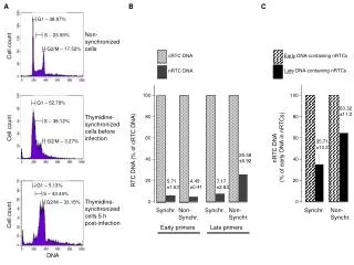
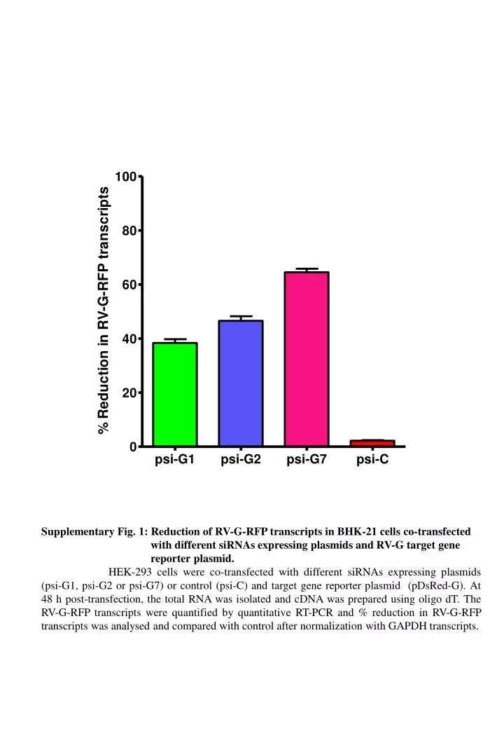
Supplementary Fig. 1: Reduction of RV-G-RFP transcripts in BHK-21 cells co-transfected with different siRNAs expressing plasmids and RV-G target gene reporter plasmid. HEK-293 cells were co-transfected with different siRNAs expressing plasmids (psi-G1, psi-G2 or psi-G7) or control (psi-C) and target gene reporter plasmid (pDsRed-G). At 48 h post-transfection, the total RNA was isolated and cDNA was prepared using oligodT. The RV-G-RFP transcripts were quantified by quantitative RT-PCR and % reduction in RV-G-RFP transcripts was analysed and compared with control after normalization with GAPDH transcripts.
FITC psi-G2 psi-G1 psi-G7 DAPI psi-G2 psi-G1 psi-G7 FITC Mock psi-C DAPI Mock psi-C Supplementary Fig. 2: Evaluation of siRNAs to inhibit RV multiplication in BHK-21 cells treated with different siRNAs expressing plasmids targeting RV-G gene. BHK-21 cells were treated with different siRNAs expressing (psi-G1, psi-G2 and psi-G7) or control (psi-C) plasmids by transfection and challenged with 0.01 MOI of RV-PV-11 strain. At 48 h post-challenge, presence of RV in infected cells was detected by staining with anti-rabies nucleocapsid FITC-labelled antibody and counterstained with DAPI. The cells with green fluorescence indicated RV-infection (200X).
Supplementary Fig. 3: Evaluation of inhibition of RV production in BHK-21 cells treated with different siRNAs expressing plasmids targeting RV-G gene. BHK-21 cells were treated with different siRNAs expressing plasmids (psi-G1, psi-G2, psi-G7 or control psi-C) by transfection and challenged with 0.01 MOI of RV-PV-11 strain. At 48 h post-challenge, the infected cell culture supernatant was harvested and amount of RV in the harvest was quantified using rabies fluorescent foci unit (ffu) assay. The percent reduction in RV titer was determined in comparison with control.