Download
1 / 19
210 likes | 331 Views
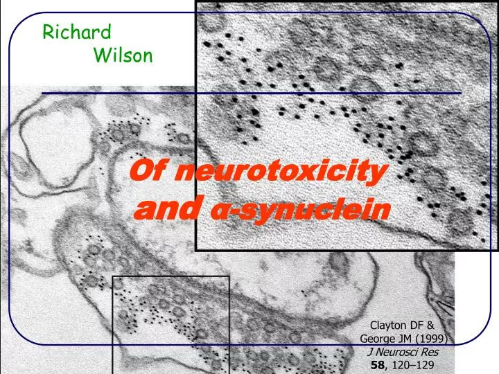
Of neurotoxicity and α -synuclein. Richard Wilson. Clayton DF & George JM (1999) J Neurosci Res 58 , 120–129. Presentation outline. Motivation for the miniproject Neurons and disease The pore hypothesis The study Results Discussion. Motivation. No – not Catholic fervour…

E N D
Of neurotoxicityandα-synuclein Richard Wilson Clayton DF & George JM (1999) J Neurosci Res 58, 120–129
Presentation outline • Motivation for the miniproject • Neurons and disease • The pore hypothesis • The study • Results • Discussion
Motivation • No – not Catholic fervour… • but a desire to combat Parkinson’s disease (PD) • Second most common neurological disease in the elderly (in the western world) • PD symptoms • Progressive loss of motor function: difficulty in initiating movements, rigidity, staggering, resting tremor • No cure available
Neurons and PD • Neurons are specialised cells forming the nervous system • All neurons produce neurotransmitters • Dopaminergic neurons produce dopamine • Dopamine is essential for motor control • PD results from destruction of dopaminergic neurons • But what is killing these cells?
Lewy bodies • Neurons of PD patients contain characteristic inclusions called Lewy bodies • visible under the light microscope http://medweb.bham.ac.uk/
The cause of PD? • Lewy bodies (LBs) are largely composed of fibrillar aggregates of the protein α-synuclein • α-Synuclein in its normal role (its native form) is unstructured and not aggregated • This has prompted research into how its structure is related to toxicity • Initially LBs thought to be toxic • Now believed that LBs are at least benign if not a protective response to the disease • Protofibrillar α-synuclein: an intermediate form between native and fibrillar is the suspect
Pore hypothesis • Volles & Lansbury (2003) proposed that pore-like protofibrils may puncture the cell membrane causing leakage of vital molecules and cell death 250 nm square Atomic force microscopy image Volles MJ & Lansbury PT (2003) Biochemistry 42 (26), 7871-7878 Goldberg MA & Lansbury PT (2000) Nature Cell Biology 2, 115-119
This study • Aim: to test the neurotoxicity of various forms of α-synuclein • Method • Induce protein aggregation • Culture cells • Add protein to cells • Test for toxicity • The experiments were performed in vitro (actually in plastic) using • artificially synthesised (recombinant) α-synuclein • B104 rat neuroblastoma cells (from central nervous system)
1) Inducing protein aggregation • α-Synuclein incubated according to published conditions (Hoyer et al 2002) • 250 μM protein incubated at 37 degC with shaking • at pH 4: 20 μM citric acid buffer, sample taken at 1, 3 and 6 hours • at pH 7: 10 μM phosphate buffer, sample taken at 24, 72 and 96 hours • Several techniques were used to check structural changes including electron microscopy Hoyer W, Antony T, Cherny D, Heim G, Jovin TM & Subramaniam V (2002) J Mol Biol322, 383-393
Incubated protein structure pH 4 protein – amorphous aggregates pH 7 protein – protofibrillar aggregates
2) Culturing cells • Cells introduced to rows of 6 wells in 96-well plates • 4 plates of cells cultured in medium with antibiotics for 4 days at 37 degC in an incubator: • 2 plates with serum to give undifferentiated cells • 2 without serum to give differentiated cells (more like adult neurons) A 96-well plate
3) Adding protein to cells • For each plate, 3 control rows • medium only • cells plus buffer • cells plus Tween 20% (kills cells) • and 3 experimental rows • cells plus 1 μM protein • cells plus 5 μM protein • cells plus 10 μM protein • Plates incubated for 24 hours at 37 degC in 10% CO2 humidified atmosphere
4) Toxicity testing: MTT assay • 3-(4,5-dimethylthiazol-2-yl)-2,5-diphenyltetrazolium bromide (MTT) was added to the wells • Plates incubated for 4 hours • Healthy cells reduce pale yellow MTT to dark blue formazan • Lysis solution (15% SDS/50% N,N-dimethylformamide) added to wells to release the formazan from the cells • Plates read by automatic plate reader (absorbance measured at 570 nm) • Resulting data averaged and normalised to the positive control (cells plus buffer) • Results plotted as bar charts with standard deviation error bars
Results – pH 4 proteins No toxicity(!)
Results – pH 7 proteins Enhanced function! Toxicity?
Discussion • The results are unexpected since • previous studies* found both native and fibrillised α-synuclein to be neurotoxic • And surprising as • the function of undifferentiated cells was enhanced by α-synuclein protofibrils • Enhancement has not been observed before • However, the results are tentative because of the lack of replication *El-Agnaf et al (1998) FEBS Letters 440, 71-75; Sung et al (2001) J Biol Chem276, 27441–27448
Implications for PD theory • If these tentative results were confirmed, then it is clear that • protofibrils don’t puncture the cell membrane • But, of course, protofibrils may attack vesicles or mitochondrial membranes • One other possibility is that B104 cells are not a good model for PD…
Acknowledgments • Biological Sciences • Teresa Pinheiro, supervisor • Bruno Correia, mentor • Narinder Sanghera, cell wizard • EPSRC, essential funding • MOAC: thanks for your support