Purpose
Results. Abstract. PET/CT 영상에서 금속 치과 재료에 의한 인공물에 관한 연구. 연세의료원 세브란스병원 핵의학과. 반영각 , 박훈희 , 남궁혁 , 조석원 , 강신창 , 임한상 , 이창호. Purpose

Purpose
E N D
Presentation Transcript
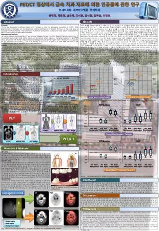
Results Abstract PET/CT 영상에서 금속 치과 재료에 의한 인공물에 관한 연구 연세의료원 세브란스병원 핵의학과 반영각, 박훈희, 남궁혁, 조석원, 강신창, 임한상, 이창호 Purpose The X-ray attenuation coefficient based on CT images is used for attenuation correction in PET/CT. The polychromatic X-ray beam can introduce beam-hardening artifact on CT images. The aims of the study were to evaluate the effect of dental metal prostheses in phantom and patients on apparent tracer activity measured with PET/CT when using CT attenuation correction. Materials and Methods 40 normal patients (mean age 54±12) was scanned between Jan and Feb 2010. Region of interest were drawn in non-artifact region, bright steak artifact region and dark streak artifact region on the same transaxial CT and PET slices. 20 patients with dental metal prostheses and 20 patients with dental implant were evaluated the change rate of CT Number and SUVmean of oral cavity in PET/CT. A paired t-test was performed to compare the ratio and the difference of the calculated values. Results In patients with dental metal prostheses, CT Number was reduced 1102.01% in the non-steak artifact region than the bright streak artifact region whereas was increased 1866.77% in the non-steak artifact region than the dark streak artifact region. SUVmean was reduced 19.64% (p <0.05) in the non-steak artifact region than the bright streak artifact region whereas was increased 90.17% (p> 0.05) in the non-steak artifact region than the dark streak artifact region. In patients with dental implant, CT Number was reduced 1544.88% in the non-steak artifact region than the bright streak artifact region whereas was increased 1796% in the non-steak artifact region than the dark streak artifact region. SUVmean was increased 19.1% (p <0.05) in the non-steak artifact region than the bright streak artifact region whereas was increased 96.62% (p> 0.05) in the non-steak artifact region than the dark streak artifact region. Conclusion When CT is used for attenuation correction in patients with dental metal prostheses, 19.1% reduced SUVmean is anticipated in the dark streak artifact region on CT images. The dark streak artifacts of CT by Dental metal prostheses may cause false negative finding in PET/CT. We recommend that the non-attenuation-corrected PET images also be evaluated for clinical use. 치아 보철을 매식한 환자의CT 에서CT 값 (CT Number) 이 인공물의 영향이 없는 부분 (Non-streak artifact region)은 최고 65.28±11, 평균46.21±1, 어둡게 인공물이 나타난 부분 (Dark streak artifact region) 은 최고 -333.5±135, 평균-463.07±211 이고, 밝게 인공물이 나타난 부분(Bright streak artifact region) 은 최고 635±221, 평균 515.57±207 이었다. 인공물의 영향이 없는 부분 (Non-streak artifact region)은 어둡게 인공물이 나타난 부분 (Dark streak artifact region) 에 비해약 1102.01 % 감소되었고, 밝게 인공물이 나타난 부분(Bright streak artifact region)은1866.77 % 증가하였다. PET 검사에서 표준섭취계수 (SUVmean) 가 인공물의 영향이 없는 부분 (Non-streak artifact region)은 최고 0.79±0.15, 평균0.8±0.15, 어둡게 인공물이 나타난 부분 (Dark streak artifact region) 은 최고 0.76±0.43, 평균 0.76±0.43이고, 밝게 인공물이 나타난 부분(Bright streak artifact region) 은 최고 1.54±0.55, 평균 1.52±0.56 이었다. 인공물의 영향이 없는 부분 (Non-streak artifact region) 에 비해 어둡게 인공물이 나타난 부분 (Dark streak artifact region) 은약19.64 % (p<0.05) 감소되었고, 밝게 인공물이 나타난 부분 (Bright streak artifact region) 은90.17 % (p>0.05) 증가하였다. 치아 임플란트를 매식한 환자의CT 에서CT 값 (CT Number) 이 인공물의 영향이 없는 부분 (Non-streak artifact region)은 최고 62.25±13, 평균44.83±13, 어둡게 인공물이 나타난 부분 (Dark streak artifact region) 은 최고 -522.25±333, 평균-649.58±363 이고, 밝게 인공물이 나타난 부분(Bright streak artifact region) 은 최고 1021±372, 평균 801.08±348 이었다. 인공물의 영향이 없는 부분 (Non-streak artifact region)은 어둡게 인공물이 나타난 부분 (Dark streak artifact region) 에 비해약 1548.88 %감소되었고, 밝게 인공물이 나타난 부분(Bright streak artifact region)은1796 % % 증가하였다. PET 검사에서 표준섭취계수 (SUVmean) 가 인공물의 영향이 없는 부분 (Non-streak artifact region)은 최고 0.75±0.16, 평균0.74±0.15, 어둡게 인공물이 나타난 부분 (Dark streak artifact region) 은 최고 1±0.43, 평균 0.99±0.42이고, 밝게 인공물이 나타난 부분(Bright streak artifact region) 은 최고 1.5±0.46, 평균 1.45±0.4 이었다. 인공물의 영향이 없는 부분 (Non-streak artifact region) 에 비해 어둡게 인공물이 나타난 부분 (Dark streak artifact region) 은약19.1 % (p<0.05) 감소되었고, 밝게 인공물이 나타난 부분 (Bright streak artifact region) 은96.62 % (p>0.05) 증가하였다(Fig. 5, Fig. 6). Introduction 최근 종양학에서PET 영상은 PET 장비보다 PET/CT 장비의 개발로 빠르게 대체되고 있다 (Fig. 1). 그것은 기존PET 장비의 68Ge 을 이용한 감쇄보정보다 PET/CT 검사에서CT 기반의 감쇄보정은 기존의 감쇄보정 방법보다 더 신속하게 검사가 진행되기 때문이다(Fig. 2). 이러한PET 영상장비의 발전으로 전신PET영상의 검사시간은30%-40% 감소 되었고, 따라서 검사할 수 있는 환자 수 가 기존의 PET 에 비해 많이 늘어났다. 게다가CT 영상을 획득함으로써PET 영상과CT 영상의 동위상 정보를 제공하여 정확한 해부학적, 기능적 영상을 동시에 얻을 수 있다. 그러나 이러한 장점에도 불구하고CT 기반의PET/CT 감쇄보정은 단점을 가지고 있다. PET/CT 검사에서는 CT 영상의 인공물로 인해 PET/CT 의 감쇄보정에 영향이 있을 수 있는 것이다. CT 기반의 감쇄보정 영상은X-ray 의 감쇄 차이를 영상으로 획득하여PET 영상의 감쇄된 영상을 보정하는데, CT 영상은X-ray 의 특성상 선속 경화 Dental Metal Implants Patients Dental Metal Prostheses Patients Fig. 5 CT Numbers and SUV were measured scatter plots graph in patients with implants and prostheses. Fig. 1 PET & PET/CT Change rate in Korea. Max Mean Background Max Mean Background Max Mean Background Max Mean Background Max Mean Dark region Max Mean Dark region Max Mean Dark region Max Mean Dark region Max Mean Bright region Max Mean Bright region Max Mean Bright region Max Mean Bright region 인공물로 인해 영상에 영향을 미칠 수 있고, PET/CT 영상에서 선속이 경화된 CT 영상이 감쇄보정에 이용되어 PET 영상에서도 영향을 미칠 수 있다. 이러한 현상은 인공 심장 박동기, 금속 치과 재료, 정형외과에서 쓰는 인공관절 등이 CT 영상에서 X-ray 선속 경화로 인해 인공물이 발생하고, 이것은 PET 영상에서 영향을 미치고 있다. 특히, 구강 내에서의 금속 치과 재료로 인한 인공물에 의해 PET/CT 검사에서 두경부의 정확한 판독이 어렵다. 그래서 본 연구는 치과재료에 의한 인공물이 PET/CT 영상에 미치는 영향에 대해 알아보고자 하였다. CT Number SUV Materials & Methods 67Ge AC Non-AC PET PET Image Dental Metal Implants Patients CT Number SUV CT AC Non-AC PET PET Image Conclusion Fig. 2 PET and PET/CT Attenuation correction image process. 환자 정보 2009년 9월부터 2010년 1월까지 본원을 내원한 구강 내질환이 없는 치아에 치과 보철을 매식한 환자 20 명과 치과 임플란트를 매식한 환자 20 명을 대상으로 하였으며, 남자 22명 여자 18명 이었고, 평균 연 령은 54±12세였다(Fig. 3). 영상획득방법 양전자방출단층촬영은 환자에게 방사성동위원소주사 전 500-1000cc 정도의 충분한 수분을 섭취하게 한 후 18F-FDG (5~9 mCi) 를말초혈관에 정맥 주사하였으며, 측정한 선량을 모두 인체에 주입하기 위하여 3-way stop cock 을이용하여 생리식염수 100cc 를 30 초 간 천천히 정맥 주사 하였다. 환자는 주사 전 최소 8 시간 이상 금식하였고, 검사직전 혈당은 모두6.69 mmol/l (120 mg/dl) 이하였다. 주사 후에 근육 섭취를 막기 위하여 40 – 50 분간 움직임을 제제하였다. 주사 60 분 후에 전신 검사를 시행 하였으며, 투과스캔(transmission scan)을저선량 CT (CTAC) 영상 획득 후 방출스캔(emission scan)을 Bed 당 2분 30초 획득하였다. 영상분석방법 치아에 치과 보철을 매식한 환자20명과 치과 임플란트를 매식한 환자20명을 대상으로 같은 단층상의PET/CT 영상에서 인공물의 영향이 없는 부분, 어둡게 인공물이 나타난 부분, 밝게 인공물이 나타난 부분, 이렇게 3군데의 관심영역을 설정하여 CT 영상의 CT Number 와PET 영상의 표준섭취계수 변화를 측정하였다(Fig. 4). 통계분석은Paired T-test 를 이 용하였다. A Discussion Dental Metal Prostheses Patients B References Fig. 6 CT Numbers and SUV were measured box plots graph in patients with implants and prostheses. PET/CT 검사의 CT 기반 보정 영상 구현 시 금속 치과 재료로 인해 CT 값 (CT Number) 에 영향을 주어 PET 영상에서도 표준섭취계수 (SUVmean) 에 영향이 미치는 것을 볼 수 있었다. 그리고 치아 임플란트 보다 치아 보철에서 과보정이 된 것을 볼 수 있었다. 특히, 치아 보철에서 표준섭취계수 (SUVmean) 가 어둡게 인공물이 나타난 부분에서 19.64 % 감소되어 나타난 것을 확인 할 수 있어 위음성으로 오인할 가능성이 있다. 그러므로 금속 치아 보철이나 임플란트를 시행한 환자들의 PET/CT 검사 시 구강 내 병변이 의심 된다면, CT 영상으로 보정이 되지 않은 무보정 (Non attenuation correction) PET 영상을 확인하는 것이 정확한 진단에 도움을 줄 것으로 사료된다. Fig. 3 A: Implant and B: Prostheses with patients. 우리가 알고 있듯이 CT 값에따라서 PET/CT 영상에서표준섭취계수에영향을 미치는 것을 알 수 있다. 대부분은 CT 값이 높게평가 된 영역에서 표준섭취계수도 높게 평가되는 현상을 보여 주고 있다. 하지만,CT 값이낮게 평가 된 부분은 표준섭취계수가 많은 차이로 낮게 평가 되지는 않는다. 그래서, 암 환자에 있어서 영상진단을 할 때 확실한 정보를 제공하기 위하여 CT 로보정되지 않은 즉, CT 값에영향을 받지 않은 무보정 PET (Non-attenuation correction PET image) 영상을제공하는 것이 확실한 진단을 내리는데 도움이 될 것이다. • Waheeda Sureshbabu, Osama Mawlaqwi. PET/CT Imaging Artifacts. J Nucl Med Technol 2005; 33:156- • 161. • 2. Hiroaki Shimamoto, Naoya Lalimoto, Kouichi Fujino, Seiki Hamada, Eku Shimosegawa, Shumei Murakami, etc. Metalic artifacts cause by dental metal prostheses on PET images: a PET/CT Phantom study using different PET/CT scanners. Ann Nucl Med 2009; 23:443-449. • 3. Sandra J. Rosenbaum, Thomas Lind, Gerald Antoch, Andreas Bockisch. False-Positive FDG PET Uptake- • the Role of PET/CT. Eur Radiol 2006; 16:1054-1064. • 4. Ehanb M. Kamel, Cyrill Burger, Alfred Buck, Gustav K. von Schulthess, Gerhard W. Goerres. Impact of Metalic dental implants on CT-based attenuation correction in a combined PET/CT scanner. Eur Radiol 2003; • 13:724-728. Fig. 4 ROI s were designed PET/CT images in implants and prostheses with patients