Download
1 / 64
651 likes | 1.96k Views
Learn about ankle joint anatomy, ligaments, types of injuries, radiological views for diagnosis, classifications, syndesmotic injuries, management of fractures, complications, and talus fractures. Understand the treatment options and different types of ankle fractures.

E N D
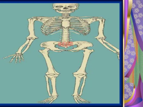
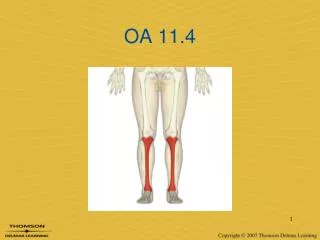
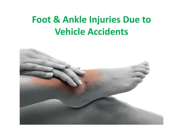
INTRODUCTION • Ankle injury refers to disruption of any component or components of the ankle joint following trauma. • Ankle injuries occur frequently, and have high propensity for complications.
ANATOMY • Ankle joint is a synovial joint of hinge variety
Bony mortise- quadrilateral shape • Posterolateral position of fibula • Ligaments 3 groups -Lateral -Medial -Syndesmotic
ANKLE JOINT IS SUPPORTED BY • Fibrous capsule • Deltoid ligament A. Superficial a. Anterior- Tibionavicular b. Middle- Tibiocalcanean c. Posterior- Posterior tibiotalar B. Deep : Anterior-Tibiotalar
Lateral ligament • Anterior- Talofibular • Posterior- Talofibular • Calcaneofibular
SYNDESMOTIC LIGAMENTS • Ant inf tibio fib • Supf post tibio fib • Deep post tibio fib • Interosseous lig
ACUTE LIGAMENTOUS INJURY • Type I sprain- minor • Type II sprain - incomplete • Type III sprain - complete
TREATMENT • LIGAMENT INJURY • Non-operative treatment • Achieved by RICE • Operative treatment • Indicated when problems persist after 12 weeks of treatment including physiotherapy • Associated fracture
CLASSIFICATIONS • LAUGE HANSEN
LAUGE HANSEN • Position of foot at injury- Pronation/Supination • Deforming force- Abduction/ adduction/ external rotation • Most Common mechanism of injury- SER • Most Common unstable ankle fracture variant- SER
LAUGE HANSEN • SUPINATION ADDUCTION • SUPINATION EXT ROT • PRONATION ABDUCTION • PRONATION EXT ROT • PRONATION DORSIFLEX
Maisonneuve’s fracture • High spiral oblique fracture of upper 3rd fibula with ankle PER injury
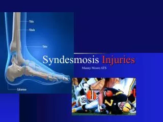
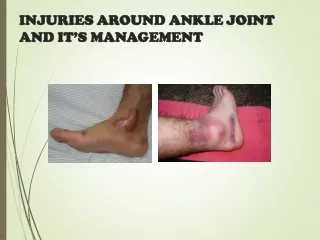
TYPES OF INJURIES • Soft tissue injuries • Ligament injuries • Lateral collateral ligament injury • Deltoid ligament injury • Syndesmotic injury • Fractures • Malleolar fractures • Pilon fractures • Physeal injuries
RADIOLOGICAL VIEWS • AP / LAT ANKLE • AP/OBLIQUE FOOT • AP MORTISE ANKLE
OTHER INVESTIGATIONS • ARTHROGRAPHY • ARTHROSCOPY • CT SCAN • MRI • BONE SCAN
AP VIEW • SYNDESMOSIS • Tibiofibular overlap<10mm • MALLEOLAR LENGTH • Talocrural angle 83+_4 deg • TALAR TILT - sup clear space- med clear space diff <2mm
What else to see in x-rays LAT MALLEOLUS • Level of fracture • Orientation of fracture • Fracture comminution MED/POST MALLEOLUS • Size • Assoc plafond # • Assoc syndesmotic injury
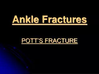
Pott’s Fracture • Fracture involving the ankle joint loosely referred to as Pott’s Fracture • First degree single malleolus fractured. • In second degree two malleoli are fractured. • In third degree there is bimalleolar fracture with a fracture of posterior part of inferior articular surface of the tibia referred to as third malleolus. (Tri Malleolar fracture)
MANAGEMENT • RICE Definitive • Aim- restoration of complete normal anatomical alignment of ankle. • Patients if needs operation should be operated within 24hrs of injury or after one week once the swelling subsides. Undisplaced fracture medial malleolus : • Below knee POP cast for 6 weeks. • Reduction fails (may be due to soft tissue (periosteal) inter position)
Displaced: • Open reduction and internal fixation by • Cancellous screws group • Tension band wiring Fracture lateral malleolus: • Lateral Malleolus helps in length maintenance & maintenance of ankle mortice. • Hence, lateral malleolus has to be fixed internally.
TIBIAL PILON FRACTURES • Intraarticular fracture of distal tibia. • Fibula is fractured in 85% of these patients.
TIBIAL PILON FRACTURE • Plaster immobilization • Traction • Lag screw fixation • OR & IF with plates • External fixation with or without limited internal fixation If articular incongruity <2 mm and reserved for low energy injuries
COMPLICATIONS • Malunion- may result in posttraumatic arthritis and painful movements. • Nonunion of medial malleolus- commonly due to interposition of fractured periosteum between two fragments. • Repeated edema • Sudeck’s Osteodystrophy
Anatomy-parts • Head-articulate with navicular • Neck-nonarticular • Body-articulate with tibia and calcaneus • No muscular or tendinous attachment
Blood supply • Extraosseous supply • Posterior tibial a. tarsal canal a. • Anterior tibial a. sinus tarsi a • Peroneal a. sinus tarsi a. • Intraosseous supply • Talar head • Talar body -anastomosis between tarsal canal a. and tarsal sinus a.
Talar head fracture • 5~10% of all talus fracture
Talar neck fracture • Aviator’s astragalus • High energy injury, hyperdorsiflexion • 15~20% open fracture • Associated with malleloar fracture(25% of cases), medial malleolus is more common • High risk of soft tissue injury and compartment syndrome
Classification-Hawkins classification Displaced Subtalar subluxation nondisplaced Ankle dislocation (Talar body dislocation) Talonavicular dislocation
Treatment • Hawkins type I • 4~6 weeks of no weightbearing in a short leg cast walking cast for 1~2 months • Percutaneous screw fixation
Treatment • Hawkins type II • Orthopaedic emergency: traction and plantar flexion by manipulation anatomic reduction(50%) treated as type I • Open reduction: screw placed across the neck fracture
Treatment • Hawkins type III • ORIF and Skeletal traction through the calcaenus • Open fracture (> type III) :talar body excision followed By primary tibiocalcaneal or Blair-type arthrodesis • Hawkins type IV • Rare injury • As type II
Complication • Skin necrosis and infection • Delayed union or nonunion • Malunion • Posttraumatic arthritis • Osteonecrosis
Anatomy • Largest, most irregularly shaped bone in foot • Large calcellous bone and multiple processes • Achilles tendon posteriorly and plantar fascia inferiorly : tuberosity • Posterior facet: talar lateral process and body • Middle facet: Sustentacular fragment (flexor hallucislongus pass) • Anterior process: cuboid
Calcaneal fracture • Classification • Essex-Lopresti --Extraarticular(25%) v.s intraarticular(75%) fracture • Sanders --CT classification of intraticular calcaneal fracture
Associated injuries • A fall from a height or high–energy mechanisms • 10% lumbar spine fracture(L1); 10% of calcaneal fracture are bilateral
↓ ↑ varus position of the tuberosity Broden’s view showing the depressed posterior facet
Intraarticular fracture(joint depression and tongue type) • Mechanism injury • Axial loading • Radiography • Loss of Bohler’s and Gissane’s angles
Intraarticular fracture Joint-depression type, in which the primary fracture line exited the bone close to the subtalar joint tongue-type, in which the primary fracture line exited the bone posteriorly
Intraarticular fracture--Treatment • Nondisplaced articular fractures • Bulky (Robert-jones) dressing: active subtalar ROM, prohibit weightbearing walking 8~12 wks later • Displaced intraarticular fracture with large fragment • ORIF
Intraarticular fracture--Treatment • Displaced intraarticular fracture with severe comminution • Increasing intraarticualr comminution leads to less satisfactory results • ORIF primary arthrodesis • Restoring the heel width and height