Download

1 / 45
450 likes | 476 Views
Explore direct and indirect methods for measuring blood pressure, sensor types, and dynamic properties of pressure sensors. Learn about physical and electrical models for catheter-sensor systems.

E N D
Blood pressure measurement (Ideal) Target: Blood pressure @ the heart chambers @ the peripheral vascular system Purpose: to help the physician to determine the functional integrity (完整) of the cardiovascular system • Methods: • Direct (invasive) techniques • 2. Indirect (noninvasive) techniques
Phonocardiography The sources of heart sounds: the variations set up by the accelerations and deccelerations of blood Function of blood circulation: oxygen, nutrient Tissues metabolic waste Tissues
組織 大動脈 下大靜脈 肺 肺 組織 Figure 7.1 The left ventricle ejects blood into the systemic circulatory system. The right ventricle ejects blood into the pulmonary circulatory system.
7.1 Direct Measurements Blood pressure sensors: Type 1) Extravascular pressure sensors Type 2) intravascular pressure sensors Types of sensor elements: Strain gage, linear-variable differential transformer, variable inductance, variable capacitance, optoelectronic, piezoelectric, semiconductor
c Diaphragm R2 R1 Rx A b ui B a Ry R4 R3 Armature C d D Ri Duo (a) Strain-gage wires (b)
c Diaphragm R2 R1 Rx A b ui B a Ry R4 R3 Armature C d D Ri Duo (a) Strain-gage wires (b) Figure 2.2 (a) Unbonded strain-gage pressure sensor. The diaphragm is directly coupled by an armature to an unbonded strain-gage system. With increasing pressure, the strain on gage pair B and C is increased, while that on gage pair A and D is decreased. (b) Wheatstone bridge with four active elements. R1 = A, R2 = B, R3 = D, and R4 = C when the unbonded strain gage is connected for translation motion. Resistor Ry and potentiometer Rx are used to initially balance the bridge. vi is the applied voltage and Dv0 is the output voltage on a voltmeter or similar device with an internal resistance of Ri.
Figure 2.2 c Diaphragm R2 R1 Rx A b ui B a Ry R4 R3 Armature C d D Ri Duo (a) Strain-gage wires (b) Figure 2.2 (a) Unbonded strain-gage pressure sensor. The diaphragm is directly coupled by an armature to an unbonded strain-gage system. With increasing pressure, the strain on gage pair B and C is increased, while that on gage pair A and D is decreased. (b) Wheatstone bridge with four active elements. R1 = A, R2 = B, R3 = D, and R4 = C when the unbonded strain gage is connected for translation motion. Resistor Ry and potentiometer Rx are used to initially balance the bridge. vi is the applied voltage and Dv0 is the output voltage on a voltmeter or similar device with an internal resistance of Ri.
c Diaphragm R2 R1 Rx A b ui B a Ry R4 R3 Armature C d D Ri Duo (a) Strain-gage wires (b) Figure 2.2 (a) Unbonded strain-gage pressure sensor. The diaphragm is directly coupled by an armature to an unbonded strain-gage system. With increasing pressure, the strain on gage pair B and C is increased, while that on gage pair A and D is decreased. (b) Wheatstone bridge with four active elements. R1 = A, R2 = B, R3 = D, and R4 = C when the unbonded strain gage is connected for translation motion. Resistor Ry and potentiometer Rx are used to initially balance the bridge. vi is the applied voltage and Dv0 is the output voltage on a voltmeter or similar device with an internal resistance of Ri.
Flush solution under pressure Sensing port Roller clamp Sample and transducer zero stopcock Electrical connector Disposable pressure transducer with an integral flush device Figure 7.3 Extravascular pressure-sensor system A catheter couples a flush solution (heparinized saline) through a disposable pressure sensor with an integral flush device to the sensing port. The three-way stopcock is used to take blood samples and zero the pressure sensor.
7.3 Dynamic properties of the pressure sensor Static pressure Blood pressure (dynamic)
Properties of liquid-filled catheter sensor: Pressure (P), pascal = N/m2 Voluminal flow rate (F), m3/s Liquid volume, m3 Voltage (V), V Current (I), C/s = A Electric charge, coulomb
△P I +△V- F
Physical model & Analogous electrical model Sensor (a) P Diaphragm Catheter Liquid Incremental length DV Rc Lc Rc Lc Rc Lc Rs Ls (b) DV Cc Cc Cc C d = Cs DP Analogous (adj.) 類似的 Analog (adj.) 類比式的 Figure 7.7 (a) Physical model of a catheter-sensor system. (b) Analogous electric system for this catheter-sensor system. Each segment of the catheter has its own resistance Rc, inertance Lc, and compliance Cc. In addition, the sensor has resistor Rs, inertance, Ls, and compliance Cs. The compliance of the diaphragm is Cd.
Rc Poiseuille equation: a physical law that describes slow viscousincompressible flow through a constant circular cross-section
Sensor (a) P Diaphragm Catheter Liquid Incremental length DV Rc Lc Rc Lc Rc Lc Rs Ls (b) DV Cc Cc Cc C d = Cs DP Figure 7.7 (a) Physical model of a catheter-sensor system. (b) Analogous electric system for this catheter-sensor system. Each segment of the catheter has its own resistance Rc, inertance Lc, and compliance Cc. In addition, the sensor has resistor Rs, inertance, Ls, and compliance Cs. The compliance of the diaphragm is Cd.
Physical model & Analogous electrical model Sensor (a) P Diaphragm Catheter Liquid Incremental length DV Rc Lc Rc Lc Rc Lc Rs Ls (b) DV Cc Cc Cc C d = Cs DP Analogous (adj.) 類似的 Analog (adj.) 類比式的 Figure 7.7 (a) Physical model of a catheter-sensor system. (b) Analogous electric system for this catheter-sensor system. Each segment of the catheter has its own resistance Rc, inertance Lc, and compliance Cc. In addition, the sensor has resistor Rs, inertance, Ls, and compliance Cs. The compliance of the diaphragm is Cd.
Catheter liquid inertia Catheter liquid resistance Sensor diaphragm compliance (a) Lc Rc Lcd Rcd uo (t) ui (t) C Cd b (b) Lc Rc uo (t) Cb Cd ui (t) (c) Figure 7.8 (a) Simplified analogous circuit. Compliance of the sensor diaphragm is larger than compliance of catheter or sensor cavity for a bubble-free, noncompliant catheter. The resistance and inertance of the catheter are larger than those of the sensor, because the catheter has longer length and smaller diameter. (b) Analogous circuit for catheter-sensor system with a bubble in the catheter. Catheter properties proximal to the bubble are inertance Lc and resistance Rc. Catheter properties distal to the bubble are Lcd and Rcd. Compliance of the diaphragm is Cd; Compliance of the bubble is Cb. (c) Simplified analogous circuit for catheter-sensor system with a bubble in the catheter, assuming that Lcd and Rcd are negligible with respect to Rc and Lc.
Example 7.1 Sensor (a) P Diaphragm Catheter Liquid Incremental length DV Rc Lc Rc Lc Rc Lc Rs Ls (b) DV Cc Cc Cc C d = Cs DP Pin (real) Pout (measured)
fn = 91 Hz = 0.033 10 fn = 22 Hz = 0.137 1.0 No bubble uo (jw) ui (jw) Bubble 0.1 0.01 0.01 1 2 0.02 0.04 0.06 0.22 0.4 0.6 10 0.1 4 6 8 0.91 f / fn Figure 7.9 Frequency-response curves for catheter-sensor system with and without bubbles. Natural frequency decreases from 91 Hz to 22 Hz and damping ratio increases from 0.033 to 0.137 with the bubble present.
Sensor (a) P Diaphragm Catheter Liquid Incremental length DV Rc Lc Rc Lc Rc Lc Rs Ls (b) DV Cc Cc Cc C d = Cs DP
Figure 7.10 Transient-response technique for testing a pressure-sensor-catheter-sensor system. Surgical glove Three- way stopcock Match O-ring Air Saline Rubber washer Sphygmomanometer bulb
Figure 7.11Pressure-sensor transient response Negative-step input pressure is recorded on the top channel; the bottom channel is sensor response for a Statham P23Gb sensor connected to a 31-cm needle (0.495 mm ID). (From I. T. Gabe, "Pressure Measurement in Experimental Physiology," in D. H. Bergel, ed., Cardiovascular Fluid Dynamics, vol I, New York: Academic Press, 1972.)
Figure 7.12 A sinusoidal pressure-generator test system A low-frequency sine generator drives an underwater-speaker system that is coupled to the catheter of the pressure sensor under test. An "ideal" pressure sensor, with a frequency response from 0 to 100 Hz, is connected directly to the test chamber housing and monitors input pressure. Pressure sensor "Ideal“ sensor Catheter Saline Underwater speaker Low-frequency sine generator
7.6 Bandwidth requirements for measuring blood pressure May ignore harmonics of the blood pressure waveform higher than the tenth. e.g.: 20 Hz bandwidth would be enough for a heart rate of 120 bpm (or 2 Hz) Measurements of the derivative of the pressure signal increase the bandwidth requirements.
7.7 Typical pressure-waveform distortion Overdamping: due to air bubbles or blood clots due to pinching the catheter Catheter whip (猛然挪動): The catheter is bent and whippedin a region of high pulsatile flow
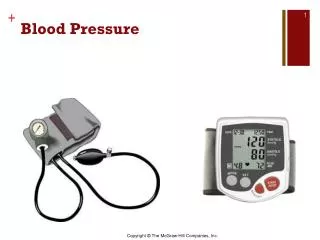
7.13 Indirect Measurements of Blood Pressure A rubber bulb for inflation of the cuff (橡膠球)(充氣) An inflatable cuff for occlusion of the blood vessel A mercury for detection of pressure
Figure 7.20 Typical indirect blood-pressure measurement system The sphygmomanometer cuff is inflated by a hand bulb to pressures above the systolic level. Pressure is then slowly released, and blood flow under the cuff is monitored by a microphone or stethoscope placed over a downstream artery. The first Korotkoff sound detected indicates systolic pressure, whereas the transition from muffling to silence brackets diastolic pressure. (From R. F. Rushmer, Cardiovascular Dynamics, 3rd ed., 1970. Philadelphia: W. B. Saunders Co. Used with permission.)