Download

1 / 36
370 likes | 617 Views
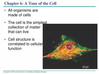
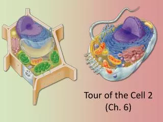
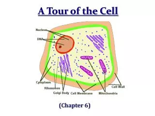
Tour of the Cell 1 (Ch. 6). Dead White Men Who Discovered (and were made of) Cells:. Anton Van Leeuwenhoek. Robert Hooke. Where the Magic Happened. The size range of cells. 10 m. Human height. 1 m. Length of some nerve and muscle cells. 0.1 m. Unaided eye. Chicken egg. 1 cm.

E N D
Dead White Men Who Discovered (and were made of) Cells: Anton Van Leeuwenhoek Robert Hooke
The size range of cells 10 m Human height 1 m Length of some nerve and muscle cells 0.1 m Unaided eye Chicken egg 1 cm Frog egg 1 mm 100 µm Light microscope Most plant and animal cells 10 µm Nucleus nucleus Most bacteria Most bacteria Mitochondrion 1 µm Electron microscope Smallest bacteria 100 nm Viruses Ribosomes 10 nm Proteins Lipids Measurements 1 centimeter (cm) = 102 meter (m) = 0.4 inch 1 millimeter (mm) = 10–3m 1 micrometer (µm) = 10–3 mm = 106m 1 nanometer (nm) = 10–3 µm = 10 9 m 1 nm Small molecules Atoms 0.1 nm
Virus Bacterium Animalcell Animal cell nucleus 0.25 m Comparing the size of a virus, a bacterium, and an animal cell While we’re on the topic of size...
Surface area increases while total volume remains constant 5 1 1 Total surface area (height width number of sides number of boxes) 6 150 750 Total volume (height width length number of boxes) 125 125 1 Surface-to-volume ratio (surface area volume) 6 12 6 Why Cells Are So Small: The SA:V Ratio Diffusion: The movement of molecules from an area of high concentration to low concentration.
In the original experiments, the researchers used microscopy to identify the organelles in each pellet, establishing a baseline for further experiments. In the next series of experiments, researchers used biochemical methods to determine the metabolic functions associated with each type of organelle. Researchers currently use cell fractionation to isolate particular organelles in order to study further details of their function. APPLICATION RESULTS TECHNIQUE Cell Fractionation Cell fractionation is used to isolate (fractionate) cell components, based on size and density. First, cells are homogenized in a blender to break them up. The resulting mixture (cell homogenate) is then centrifuged at various speeds and durations to fractionate the cell components, forming a series of pellets. Homogenization Tissue cells 1000 g (1000 times the force of gravity) 10 min Homogenate Differential centrifugation Supernatant poured into next tube 20,000 g 20 min 80,000 g 60 min Pellet rich in nuclei and cellular debris 150,000 g 3 hr Pellet rich in mitochondria (and chloro- plasts if cells are from a plant) Pellet rich in “microsomes” (pieces of plasma mem- branes and cells’ internal membranes) Pellet rich in ribosomes
Types of cells Prokaryotebacteria cells - no organelles - organelles Eukaryoteanimal cells Eukaryoteplant cells
Why organelles? • Specialized structures • specialized functions • Containers • Compartments = different local environments • pH, concentration differences • distinct & incompatible functions • lysosome & its digestive enzymes • Membranes as sites for chemical reactions • Unique lipids & proteins • embedded enzymes & reaction centers • chloroplasts & mitochondria
Cells gotta work to live! • make proteins • proteins control everycell function • make energy • for daily life • for growth • make more cells • growth • repair • renewal
Proteins do all the work! proteins cells DNA Repeat after me… Proteins do all the work! organism
Cell functions • Building proteins • copy DNA • DNA -> RNA • build proteins • process proteins • Folding, modifying • Remove amino acids • Add molecules (e.g. glycoproteins) • address & transport proteins
Golgiapparatus ribosome ER Protein Synthesis • Organelles involved • nucleus • ribosomes • endoplasmic reticulum (ER) • Golgi apparatus • vesicles The Protein Assembly Line nucleus vesicles The Endomembrane System
DNA chromosome histone protein nuclear pores nuclear pore nucleolus nuclear envelope Nucleus • Function • protects DNA • Structure • nuclear envelope • double membrane • membrane fused in spots to create pores What kind of molecules need to pass through?
nuclear membrane DNA mRNA Nucleus small ribosomal subunit nuclear pore mRNA large ribosomal subunit cytoplasm 1 production of mRNA from DNA in nucleus 2 mRNA travels from nucleus to ribosome in cytoplasm through nuclear pore
large subunit small subunit ribosome Nucleolus • Function • ribosome production • build ribosome subunits from rRNA & proteins • Ribosome assembly is completed in cytoplasm rRNA & proteins nucleolus
large subunit small subunit 0.08mm Ribosomes Rough ER Smooth ER Ribosomes • Function • protein production • Structure • rRNA & protein • 2 subunits combine
Types of Ribosomes • Freeribosomes • suspended in cytosol • synthesize proteins that stay in cytosol • Boundribosomes • attached to endoplasmic reticulum • synthesize proteins for export or membranes membrane proteins
Endoplasmic Reticulum • Function • processes proteins • manufactures membrane • synthesis & hydrolysis of many compounds • Structure • membrane connected to nuclear envelope & extends throughout cell
Types of ER rough smooth
Smooth ER function • Membrane production • Metabolic processes • Lipid Synthesis • Glycogen hydrolysis (in liver) • Drug detoxification (in liver)
Membrane Factory • Build new membrane • synthesize phospholipids • ER membrane expands • buds off & transfers to other parts of cell.
Rough ER function • Produces proteins for export out of cell • protein secreting cells • packaged into transport vesicles for export Which cellshave lot of rough ER?
cisternal space polypeptide signal sequence ribosome membrane of endoplasmic reticulum mRNA cytoplasm Synthesizing proteins ribosome
secretory vesicles transport vesicles Golgi Apparatus • Function • finishes, sorts, tags & ships products • like “UPS shipping department” • ships products in vesicles • membrane sacs • “UPS trucks” Which cellshave lots of Golgi?
protein vesicle budding from rough ER migrating transport vesicle fusion of vesicle with Golgi apparatus ribosome Vesicle transport
Putting it together… nucleus cell membrane nuclear pore protein secreted rough ER vesicle ribosome proteins smooth ER transport vesicle Golgi apparatus cytoplasm The Endomembrane System
1.. In which cell would you expect to find the most smooth endoplasmic reticulum? • Muscle cell in the thigh muscle of a long-distance runner • Pancreatic cell that manufactures digestive enzymes • Macrophage (white blood cell) that engulfs bacteria • Epithelial cells lining the digestive tract • Ovarian cell that produces estrogen (a steroid hormone)
2. In which cell would you expect to find the most bound ribosomes? • Muscle cell in the thigh muscle of a long-distance runner • Pancreatic cell that manufactures digestive enzymes • Macrophage (white blood cell) that engulfs bacteria • Epithelial cells lining the digestive tract • Ovarian cell that produces estrogen (a steroid hormone)
3. Of the following, which is probably the most common route for membrane flow in the endomembrane system? • Golgi →lysosome → ER → plasma membrane • tonoplast → plasma membrane → nuclear envelope → smooth ER • nuclear envelope → lysosome → Golgi → plasma membrane • rough ER → vesicles → Golgi → plasma membrane • ER → chloroplasts → mitochondrion → cell membrane