Microcirculation
Microcirculation. Dr Mahvash Khan MBBS, MPhil. Capillaries are the sites for exchange of materials between blood and tissue cells .

Microcirculation
E N D
Presentation Transcript
Microcirculation DrMahvashKhan MBBS, MPhil
Capillaries are the sites for exchange of materials between blood and tissue cells. • The walls of the capillaries are extremely thin, constructed of single-layer of highly permeable endothelial cells. Thereforewater, cell nutrients, and cellexcreta caninterchange quickly and easily between the tissues and the circulating blood. • The peripheral circulation of the whole body has about 10 billion capillaries with a total surface area estimated to be 500 to 700 square meters.
Structure of the Capillary Wall • The wall is composed of a single layer of endothelial cells and is surroundedby a very thin basement membrane on theoutside of the capillary. The total thickness of the capillarywall is only about 0.5 micrometers. The internaldiameter of the capillary is 4 to 9 micrometers.
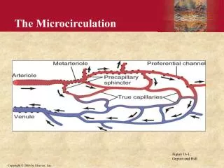
Structure of Microcirculation and Capillary System • Each nutrient artery entering an organ branches six to eight timesbefore the arteries become small enough to be called arterioles, which generallyhave internal diameter of only 10 to 15 micrometers. Then the arteriolesbranch two to five times, reaching diameterof 5 to 9 micrometers at their endswhere they supply blood to the capillaries.
The arterioles are highly muscular, and their diameter can change manyfold.Themetarterioles(the terminal arterioles) do not have a continuous muscular coat, but smooth muscle fibers encircle the vessel at intermittent points. • At the point where each capillary originates from a metarteriole, smooth muscle fibers usually encircle the capillary. This is called the precapillarysphincter. This sphincter can open and close the entrance to the capillary. • The venules are larger than the arterioles and have a much weaker muscular coat. • The pressure in the venules is much less than that in the arterioles.The venulescan contract despite the weak muscle.
Pores in the Capillary Membrane • An intercellular cleft thin-slit like curving channel that lies between adjacent endothelial cells. Each cleft is interrupted by short ridges of protein attachments that hold the endothelial cells together, but between these ridges fluid can percolate freely through the cleft. The cleft normally has a uniform spacing with a width of about 6 to 7 nanometers.
Pores in the Capillary Membrane • There are many minute vesicles called caveolae in the endothelial cells. These are formed from protein caveolins. • They are believed to play role in endocytosis.
Vasomotion • Intermittent contraction of the metarterioles and precapillarysphincters. Because of this blood does not flow continuously through the capillaries.
Regulation of Vasomotion • The most important factor found to affect the degree of opening and closing of the metarterioles and precapillarysphincters is the concentration of oxygen in the tissues.When the rate of oxygen usage by the tissue is great so that tissue oxygen concentration decreases below normal the intermittent periods of capillary blood flow occur more often, and the duration of each period of flow lasts longer, thereby allowing the capillary blood to carry increased quantities of oxygen (as well as other nutrients) to the tissues.
Exchange of water, nutrients and other substances between the blood and Interstitial fluid occurs by diffusion through the capillary membrane.
Diffusion • Diffusion results from thermal motion of the water molecules and dissolved substances in the fluid, the different molecules and ions moving first in one direction and then another, bouncing randomly in every direction.
Lipid-soluble substances can diffuse directly through the cell membranes of the capillary endothelium. • Water-Soluble, non-lipid-soluble substances diffuse only through Intercellular “Pores” in the capillary membrane.
Effect of Molecular Size on Passage Through the Pores The width of the capillary intercellular cleft pores is about 6 to 7 nanometers which is about 20 times the diameter of the water molecule which is the smallest molecule that normally passes through the capillary pores. The diameter of plasma protein molecules is slightly greater than the width of the pores. Other substances such as sodium ions, chloride ions, glucose, and urea have intermediate diameters. Therefore, the permeability of the capillary pores for different substances varies according to their molecular diameters.
Effect of Concentration Difference on Net Rate of Diffusion through the Capillary Membrane • The “net” rate of diffusion of a substance through any membrane is proportional to the concentration difference of the substance between the two sides of the membrane. The greater the difference between concentration of any given substance on the two sides of the capillary membrane, the greater the net movement of the substance in one direction through the membrane.
Interstitium and Interstitial Fluid • The spaces between the cells are collectively called interstitium and the fluid in these spaces is called interstitial fluid.
Interstitium contains • Collagen fiber bundles • Proteoglycan filaments
Tissue Gel • The fluid in the interstitiumis derived by filtration and diffusion from the capillaries. • It contains almost the same constituents as plasma except for much lower concentrations of proteins. • The interstitial fluid is entrapped mainly in the minute spaces among the proteoglycan filaments. This combination of proteoglycan filaments and fluid entrapped within them is called tissue gel.
Diffusion through the gel occurs about 95 to 99 percent as rapidly as it does through free fluid. • Because of the large number of proteoglycan filaments,itis difficult for fluid to flow easily through the tissue gel. Instead, it mainly diffuses through the gel that is, it moves molecule by molecule from one place to another by kinetic, thermal motion rather than by large numbers of molecules moving together.
Free Fluid in the Interstitium • Almost all the fluid in the interstitiumis entrapped within the tissue gel. • Occasionally small rivulets of “free” fluid small free fluid vesicles are also present, which means fluid that is free of the proteoglycan molecules and therefore can flow freely
Fluid Filtration Across Capillaries Is Determinedby Hydrostatic and Colloid Osmotic Pressures, and Capillary Filtration Coefficient • The capillary pressure (Pc), which tends to force fluid outward through the capillary membrane. • The interstitial fluid pressure (Pif), which tends to force fluid inward through the capillary membrane when Pif is positive but outward when Pifis negative. • The capillary plasma colloid osmotic pressure (Πp), which tends to cause osmosis of fluid inward through the capillary membrane. • The interstitial fluid colloid osmotic pressure (Πif), which tends to cause osmosis of fluid outward through the capillary membrane.
Net Filtration Pressure • If the sum of these forces, the net filtration pressure is positive, there will be a net fluid filtration across the capillaries. If the sum of the Starling forces is negative there will be a net fluid absorption from the interstitial into the capillaries.
Net Filtration Pressure NFP = Pc - Pif- Πp + Πif
Capillary Filtration Coefficient • The Kf is a measure of the capacity of the capillary membranes to filter water for a given NFP and is usually expressed as ml/min per mm Hg net filtration pressure.
Rate of capillary Fluid Filtration Filtration = Kf xNFP
Interstitial fluid pressure in loose subcutaneous tissue is slightly less subatmosphericaveraging about -3 mmHg.
Pumping by the Lymphatic System Is the Basic Cause of the Negative Interstitial Fluid Pressure • The lymphatic system is a “scavenger” system that removes excess fluid, excess protein molecules, debris and other matter from the tissue spaces. • Normally when fluid enters the terminal lymphatic capillaries the lymph vessel walls automatically contract for a few seconds and pump the fluid into the blood circulation. This overall process creates the slight negative pressure that has been measured for fluid in the interstitial spaces.
Plasma Colloid Osmotic Pressure • Those molecules or ions that fail to pass through the pores of a semipermeable membrane exert osmotic pressure. Because the proteins are the only dissolved constituents in the plasma and interstitial fluids that do not readily pass through the capillary pores. • About 80 per cent of the total colloid osmotic pressure of the plasma results from the albumin fraction and 20 per cent from the globulins, and almost none from the fibrinogen.
The colloid osmotic pressure of normal human plasma averages about 28 mm Hg; 19 mm of this is caused by molecular effects of the dissolved protein and 9 mm is caused by sodium, potassium, and the other cations held in the plasma by the proteins.
Interstitial Fluid Colloid Osmotic Pressure • Average interstitial fluid colloid osmotic pressure is about 8 mm Hg. Although the size of the usual capillary pore is smaller than the molecular sizes of the plasma proteins, this is not true of all the pores. Therefore small amount of plasma proteins do leak through the pores into the interstitial spaces.
Exchange of Fluid Volume Through the Capillary Membrane • The average capillary pressure at the arterial ends of the capillaries is 15 to 25 mm Hg greater than at the venous ends. Because of this difference fluid filters out of the capillaries at their arterial ends but at their venous ends fluid is reabsorbed back into the capillaries.
Analysis of the Forces Causing Filtration at the Arterial End of the Capillary
Starling Equilibrium for Capillary Exchange • Under normal conditions a state of near-equilibrium exists at the capillary membrane. The amount of fluid filtering outward from the arterial ends of capillaries equals almost exactly the fluid returned to the circulation by absorption. The slight disequilibrium that does occur accounts for the small amount of fluid that is eventually returned by way of the lymphatics.
A near-equilibrium exists between the total outward forces, 28.3 mm Hg, and the total inward force, 28.0 mm Hg. • This slight imbalance of forces, 0.3 mm Hg, causes slightly more filtration of fluid into the interstitial spaces than reabsorption. It is the fluid that must be returned to the circulation through the lymphatics..
Lymphatic System • The lymphatic system represents an accessory route through which fluid can flow from the interstitial spaces into the blood. The lymphatics can carry proteins and large particulate matter away from the tissue spaces, neither of which can be removed by absorption directly into the blood capillaries
Formation of Lymph • Lymph is derived from interstitial fluid that flows into the lymphatics. Therefore lymph has almost the same composition as the interstitial fluid.
The lymphatic system is also one of the major routes for absorption of nutrients such as fats in food. • Evenlarge particles such as bacteria canpush their way between the endothelial cells of the lymphatic capillaries and in this way enter the lymph. As the lymph passes through the lymph nodes these particles are almost entirely removed and destroyed.
Rate of Lymph Flow • The total estimated lymph flow is about 120 ml/hr or 2 to 3 liters per day.
Any factor that increases interstitial fluid pressure also increases lymph flow if the lymph vessels are functioning normally. • Elevated capillary pressure • Decreased plasma colloid osmotic pressure • Increased interstitial fluid colloid osmotic pressure • Increased permeability of the capillaries
When the interstitial fluid pressure becomes 1 or 2 millimeters greater than atmospheric pressure (greater than 0 mm Hg) lymph flow fails to rise any further at still higher pressures. This results from the fact that the increasing tissue pressure not only increases entry of fluid into the lymphatic capillaries but also compresses the outside surfaces of the larger lymphatics, thus impeding lymph flow.
When a collecting lymphatic or larger lymph vessel becomes stretched with fluid, the smooth muscle in the wall of the vessel automatically contracts. Each segment of the lymph vessel between successive valves functions as a separate automatic pump. Even slight filling of a segment causes it to contract, and the fluid is pumped into the next lymphatic segment. This fills the subsequent segment, and a few seconds later it, too, contracts, the process continuing all along the lymph vessel until the fluid is finally emptied into the blood circulation. In a very large lymph vessel such as the thoracic duct, this lymphatic pump can generate pressures as great as 50 to 100 mm Hg.
Pumping Caused by External Intermittent Compression of the Lymphatics • Contraction of surrounding skeletal muscles • Movement of the parts of the body • Pulsations of arteries adjacent to the lymphatics • Compression of the tissues by objects outside the body • The lymphatic pump becomes very active during exercise often increasing lymph flow 10- to 30-fold.
Lymphatic Capillary Pump • The terminal lymphatic capillary is also capable of pumping lymph, in addition to the lymph pumping by the larger lymph vessels. • The walls of the lymphatic capillaries are tightly adherent to the surrounding tissue cells by means of their anchoring filaments. Therefore, each time excess fluid enters the tissue and causes the tissue to swell, the anchoring filaments exert pull on the wall of the lymphatic capillary and fluid flows into the terminal lymphatic capillary through the junctions between the endothelial cells.Then, when the tissue is compressed the pressure inside the capillary increases and causes the overlapping edges of the endothelial cells to close like valves. Therefore the pressure pushes the lymph forward into the collecting lymphatic instead of backward through the cell junctions.Thelymphatic capillary endothelial cells also contain a few contractile actomyosin filaments.