Download
1 / 40
500 likes | 960 Views
Explore the devices used in external beam radiotherapy including linear accelerators, cobalt-60 units, and more. Learn about treatment planning systems and clinical objectives for optimal tumor control and tissue protection.

E N D
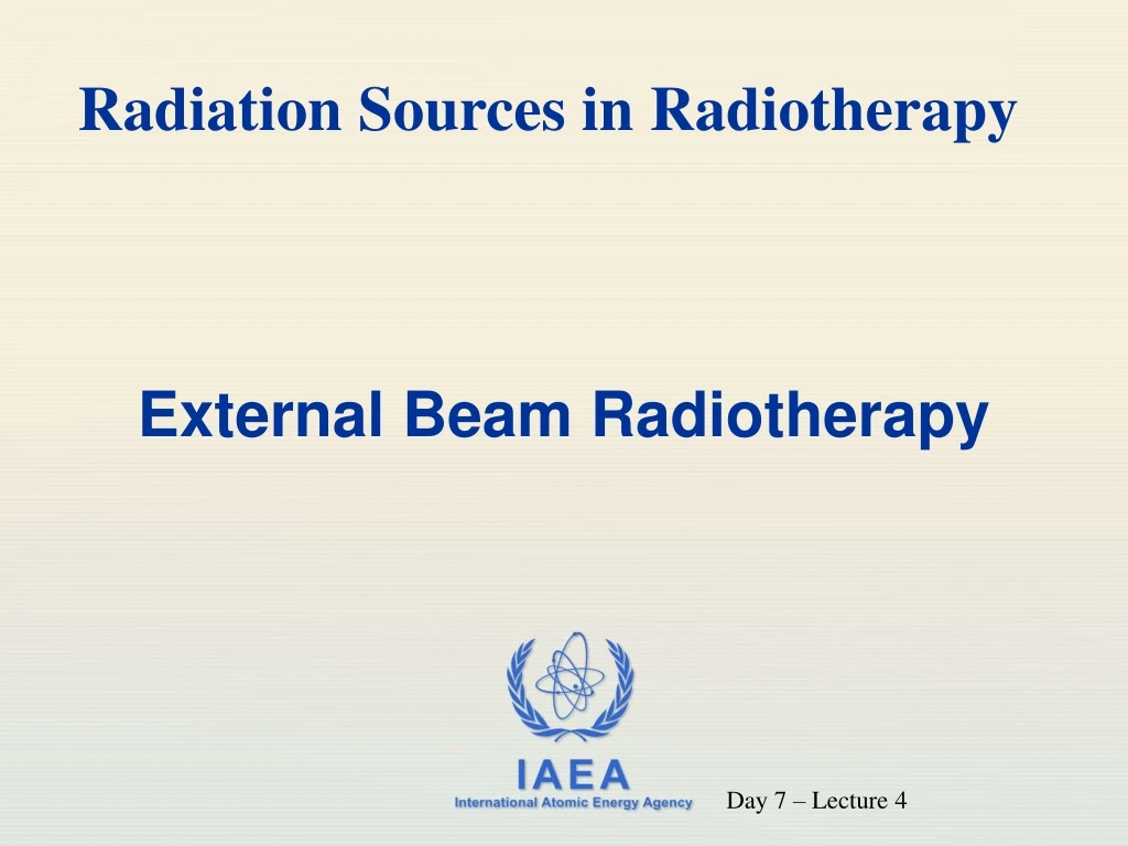
Radiation Sources in Radiotherapy External Beam Radiotherapy Day 7 – Lecture 4
Objective To become familiar with the radiation sources, devices and ancillary equipment used in external beam radiotherapy.
Contents • Treatment planning systems; • Radiotherapy simulators; • Superficial / orthovoltage units; • Cobalt-60 units including Gamma-knife; • Linear accelerators; • Computed Tomography (CT) scanners for radiotherapy; • Multileaf Collimators (MLC).
Clinical Objectives To deliver a dose and dose distribution that is adequate for tumor control but which also minimizes complications in normal tissue. Note:It is not the role of the Regulatory Body to evaluate the clinical decisions of medical practitioners authorized to prescribe radiotherapy treatments.
Treatment Planning Prescription Very important for optimization of protection in medical exposures Planning Treatment
Treatment Planning • About 1/3 of problems are directly related to treatment planning; • Problems may affect an individual patient or cohort of patients. IAEA Safety Report Series 17; 2000 “Lessons learned from accidental exposures in radiotherapy”
External Beam Equipment Therapeutic x-ray equipment operates in the range of: • 10 kVp - 150 kVp (superficial); • 150 kVp - 400 kVp (orthovoltage / deep); Radioactive sources ( γ ray equipment). • Cobalt 60 & Caesium 137 Megavoltage electron accelerators for X and electron therapy • Linear accelerator
Typical Radiation Levels • Source activity may be around 400 TBq (~10,000 Ci); Cobalt-60 teletherapy • Average radiation leakage (beam off) should not exceed 0.02 mGy/h at 1 m i.e. it would take 50 hours exposure for 1 mSv; • In general, minimize the time spent in the treatment room.
Typical radiation levels (cont) Linear accelerator turned off • There is no useful radiation beam when turned off; • However, immediately after higher energy beams (> 10 MeV) are turned off there may beinduced radioactivity but typically with very short half lives (seconds to minutes); • It is suggested that room entry be briefly delayed, especially after long exposures.
Superficial 40 kVp to 120 kVp Orthovoltage (“deep”) 150 kVp to 400 kVp Superficial and Orthovoltage x-ray equipment • treat small skin lesions to a depth of ~ 5 cm • maximum applicator size typically < 7 cm diameter • typical SSD < 30 cm • beam quality (HVL) typically 0.5 to 8 mm Al • treat skin lesions, bone metastases to a depth of ~ 20 cm • use applicators or diaphragm • SSD 30 to 60 cm • beam quality (HVL) typically 0.2 to 5 mm Cu
Superficial x-ray equipment • Interlocks prevent inappropriate combinations of kVp and filtration. • Electron contamination from the applicator can be significant.
Superficial x-ray equipment (cont) • Dose is highly dependent on source-skin distance, filtration and applicator area.
Superficial x-ray equipment (cont) Provides a range of kVp, mA and filtration Filters are used to absorb low energy photons which otherwise may unnecessarily increase skin dose.
Issues with Superficial radiotherapy • Short focus to skin distance (FSD) and hence high output and large influence of inverse square law • Calibration difficult due to strong dose gradient i.e. dose fall off and electron contamination
Issues with superficial therapy • Dose determined by a timer • on/off effects must be considered • Photon beams may be contaminated with electrons scattered from the applicator Control panel
Deep X-ray therapy (Orthovoltage) • Uses conventional X-ray tube • Energy range 150- 400 kV X-rays • Mostly used around 250 - 300 kVp • Treatment depths of around 20 mm • Applicators are used in superficial therapy X-ray tube Applicator
Deep X-ray therapy (Orthovoltage) • Penetration sufficient for palliative treatment of bone lesions relatively close to the surface (ribs, spinal cord) • Largely replaced by megavoltage treatment modalities for treatment of other lesions
Disadvantages of deep x-ray • Higher dose to bone - photoelectric absorption • Maximum dose on the surface hence higher skin dose • Treatment to a depth of only a few centimeters possible • Low energy, hence high scattered radiation and larger penumbra
Gamma ray equipment (cont) Source head and a typical source transfer mechanism
GAMMA KNIFE • The gamma knife device contains 201 cobalt-60 sources of approximately 30 curies each • It is placed in a circular array in a heavily shielded assembly. • The device aims gamma radiation through a target point in the patient's brain. • The patient wears a specialized helmetthat is surgically fixed to their skull so that the brain tumor remains stationary at target point of the gamma rays. • Therefore it is also known as the stereotacticsurgery.
Gamma Knife • uses numerous high activity 60Co sources positioned in a device so that the radiation beams converge at the specified point of treatment; • is used to treat head tumors. The Gamma Knife: Patient positioning collimator
Linear Accelerator Modern accelerators have a number of treatment options e.g. • X-rays or electrons (dual mode); • 2 X-ray energies; • 5 or more electron energies.
Linear Accelerator (cont) Concept
Linear Accelerator (cont) • is controlled by two independent integrating transmission ionization chamber systems; Radiation exposure: • one of these is designated as the primary system and should terminate the exposure at the correct number of monitor units; • these also steer the beam.
Linear Accelerator (cont) • the other system is termed the secondary system and is usually set to terminate the exposure after an additional 0.4 Gy; • most modern accelerators also have a timer which will terminate the exposure if both ionization chamber systems fail.
Linear Accelerator (cont) Complex head structure to handle multiple energies and multiple modalities.
Linear Accelerator (cont) Complex control system
Linear Accelerator (cont) Verification systems All accelerator manufacturers produce computer controlled verification systems which provide an additional check that thesettings on the accelerator console: • are correct forproper accelerator function;and • correspond exactly with theparameters determinedfor the individual patient during the treatment planning process
Linear Accelerator (cont) Rectangular (conventional) • The transmission through the collimators should be less than 2% of the primary (treatment) beam. X-ray Collimators • Multi-leaf collimators (MLC) • the transmission through the collimators should be less than 2% of the primary (treatment) beam. • The transmission between the leaves should be checked to ensure that it is less than the manufacturer’s specification.
Linear Accelerator (cont) • open sided for modern accelerators using double scattering foils or scanned beams; • enclosed for older accelerators using single scattering foils. Electron applicators may be: • Both types should be checked for leakage: • adjacent to the open beam; • on the sides of the applicators.
Linear Accelerator (cont) • should be considered if the x-ray energy is greater than 10 MV Neutrons:- Issues which need to be considered when neutrons are presents include: • neutron activation • shielding problems
General Safety Requirements • Clear indication shall be provided at the control console and in the treatment room to show when the equipment is in operation. • Dualinterlocks shall be providedon all doors to the treatment room such that opening a door will interrupt the treatment. It should only be possible to resume treatment from the control console.
General Safety Requirements Warning Signals and Signs
General Safety Requirements There shall be at least two independent “fail safe” systems for terminating the irradiation. These could be: “Fail safe” systems • two independent integrating in-beam dosemeters; • two independent timers; • an integrating dose meter and timer. Each system shall be capable of terminating the exposure.
General Safety Requirements The exposure shall be limited to the area being examined or treated by the use of collimating devices aligned with the radiation beam. Collimation Exposure rates outside the examination or treatment area due to leakage or scatter shall be kept as low as reasonably achievable.