The Larynx
The Larynx. Objectives Describe anatomical structure of larynx. Enlist the cartilages of the larynx. Define inlet of the larynx. Enlist the extrinsic and intrinsic muscles of the larynx with their nerve supply and actions. The Larynx


The Larynx
E N D
Presentation Transcript
Objectives Describe anatomical structure of larynx. Enlist the cartilages of the larynx. Define inlet of the larynx. Enlist the extrinsic and intrinsic muscles of the larynx with their nerve supply and actions.
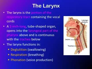
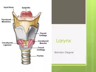
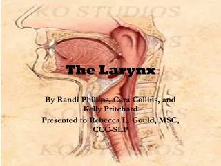
The Larynx • The larynx is an organ that provides a protective sphincter at the inlet of the air passages and is responsible for voice production. • It is situated below the tongue and hyoid bone • between the great blood vessels of the neck and • lies at the level of the fourth, fifth, and sixth cervical vertebrae . • It opens above into the laryngeal part of the pharynx, and below is continuous with the trachea. • The larynx is covered in front by the infrahyoid strap muscles and at the sides by the thyroid gland.
Cartilages of the Larynx • Thyroid cartilage: • This is the largest cartilage of the larynx (Fig. 11-98) and consists of: • Two laminae of hyaline cartilage that meet in the midline in the prominent V angle (the so-called Adam's apple). On the outer surface of each lamina is an oblique line for the attachment of muscles. • Superior cornu. • Inferior cornu.
Cricoid cartilage: • This cartilage is formed of hyaline cartilage and shaped like a signet ring, having a broad plate behind and a shallow arch in front (Fig. 11-98). It articulates with the inferior cornu of the thyroid cartilage. • Posteriorly articulates with the arytenoid cartilage. • All these joints are synovial.
Arytenoid cartilages: • There are two arytenoid cartilages, which are small and pyramid shaped and located at the back of the larynx (Fig. 11-98). • They articulate with the upper border of the lamina of the cricoid cartilage. • Each cartilage has: • An apex above that articulates with the small corniculate cartilage. • A base below that articulates with the lamina of the cricoid cartilage, and a vocal process that projects forward and gives attachment to the vocal ligament. • A muscular process that projects laterally gives attachment to the posterior and lateral cricoarytenoid muscles.
Corniculate cartilages: Two small conical-shaped cartilages articulate with the arytenoid cartilages (Fig. 11-99). They give attachment to the aryepiglottic folds. • Cuneiform cartilages: These two small rod-shaped cartilages are found in the aryepiglottic folds and serve to strengthen them (Fig. 11-99).
Epiglottis: This leaf-shaped lamina of elastic cartilage lies behind the root of the tongue (Fig. 11-98). • Its stalk is attached to the back of the thyroid cartilage. • The sides of the epiglottis are attached to the arytenoid cartilages by the aryepiglottic folds of mucous membrane.
Inlet of the Larynx • The inlet of the larynx looks backward and upward into the laryngeal part of the pharynx and is bounded: • In front by the epiglottis. • Laterally by the aryepiglottic fold. • Posteriorly by the arytenoid cartilages with the corniculate cartilages. • The cuneiform cartilage lies within aryepiglottic fold.
The piriform fossa is a recess on either side of the aryepiglottic. • Laryngeal Folds: • The vestibular fold is a fixed fold on each side of the larynx (Fig. 11-98). It is formed by mucous membrane covering the vestibular ligament and is vascular and pink in color.
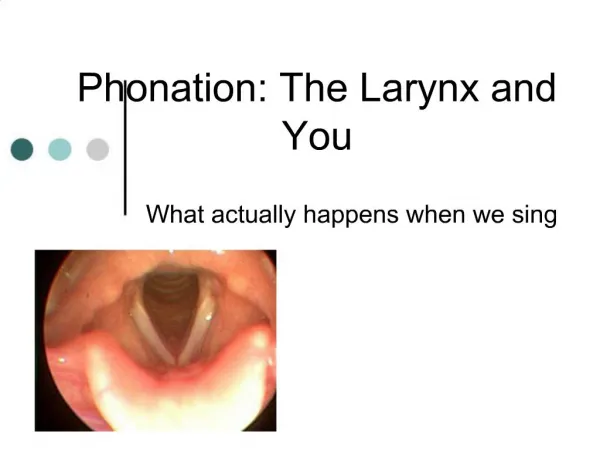
Vocal Fold • The vocal fold (Vocal Cord) is a mobile fold on each side of the larynx and is concerned with voice production. • It is formed by mucous membrane covering the vocal ligament and is avascular and white in color. The vocal fold moves with respiration and its white color is easily seen when viewed with a laryngoscope. • The gap between the vocal folds is called the rimaglottidis or glottis.
Muscles of the Larynx • The muscles of the larynx may be divided into two groups: extrinsic and intrinsic. • Extrinsic Muscles • These muscles move the larynx up and down during swallowing. Note that many of these muscles are attached to the hyoid bone, which is attached to the thyroid cartilage by the thyrohyoid membrane. It follows that movements of the hyoid bone are accompanied by movements of the larynx. • Elevation: The digastric, the stylohyoid, the mylohyoid, the geniohyoid, the stylopharyngeus, the salpingopharyngeus, and the palatopharyngeus muscles • Depression: The sternothyroid, the sternohyoid, and the omohyoid muscles
Intrinsic Muscles • Two muscles modify the laryngeal inlet (Fig. 11-99): • Narrowing the inlet: The oblique arytenoid muscle • Widening the inlet: The thyroepiglottic muscle • Five muscles move the vocal folds (cords) (Fig. 11-99): • Tensing the vocal cords: The cricothyroid muscle • Relaxing the vocal cords: The thyroarytenoid (vocalis) muscle • Adducting the vocal cords: The lateral cricoarytenoid muscle • Abducting the vocal cords: The posterior cricoarytenoid muscle • Approximates the arytenoid cartilages: The transverse arytenoid muscle
Nerve Supply of the Larynx • Sensory Nerves: • Above the vocal cords: The internal laryngeal branch of the superior laryngeal branch of the vagus. • Below the level of the vocal cords: The recurrent laryngeal nerve . • Motor Nerves: • All the intrinsic muscles of the larynx except the cricothyroid muscle are supplied by the recurrent laryngeal nerve. The cricothyroid muscle is supplied by the external laryngeal branch of the superior laryngeal branch of the vagus.
Blood Supply of the Larynx • Upper half of the larynx: The superior laryngeal branch of the superior thyroid artery • Lower half of the larynx: The inferior laryngeal branch of the inferior thyroid artery • Lymph Drainage of the Larynx • The lymph vessels drain into the deep cervical group of nodes.