Chapter 19 – Respiratory System
Chapter 19 – Respiratory System. Trachea, Bronchial Tree, Lungs. Trachea. Extends from larynx into the thoracic cavity where it splits into the left and right bronchial tree. Trachea. Structure Lined with ciliated mucous membrane

Chapter 19 – Respiratory System
E N D
Presentation Transcript
Chapter 19 – Respiratory System Trachea, Bronchial Tree, Lungs
Trachea • Extends from larynx into the thoracic cavity where it splits into the left and right bronchial tree
Trachea • Structure • Lined with ciliated mucous membrane • Membrane filters out particles and moves them up to the pharynx where they can be swallowed
Trachea • Structure • Tracheal wall contains about twenty C-shaped pieces of hyaline cartilage with smooth muscle and connective tissues between them • The cartilage prevents the trachea from collapsing and preventing air flow while the soft tissues between allow the esophagus to expand when food passes through it
Bronchial Tree • Branched airways that lead from the trachea to microscopic air sacs in the lungs
Bronchial Tree • Branches • Right and left primary bronchi arise from the trachea • These are separated by a ridge of cartilage called the carina • Secondary (or lobar) bronchi branch from the primary bronchi • Three branch on the right and two branch on the left • Tertiary (or segmental) bronchi split from the secondary bronchi • These supply a portion of the lung called the bronchopulmonary segment • Typically ten segments are in right lung and eight segments are in the left lung
Bronchial Tree • Branches • Intralobular bronchioles branch from the tertiary bronchi • These enter the basic units of the lung (called lobules) • Terminal bronchioles branch from the intralobular bronchioles • There are fifty to eighty terminal bronchioles in a lobule of the lung • Respiratory bronchioles branch from each terminal bronchiole • These have air sacs that bud from their sides and allow them to participate in gas exchange • There are typically two or more that branch from each terminal bronchiole
Bronchial Tree • Branches • Alveolar ducts branch from each respiratory bronchial • Alveolar sacs come off of the alveolar ducts • Alveoli (which are thin-walled microscopic air sacs) open to the alveolar sacs
Bronchial Tree • Structure of respiratory tubes • Bronchus • Structured similar to the trachea • C-shaped rings of cartilage are replaced by plates of cartilage that completely surround the bronchus (unlike the c-shaped rings which open posteriorly) when the bronchus enters the lung
Bronchial Tree • Structure of respiratory tubes • Smaller branches • As the branches of the respiratory tree become smaller: • The amount of cartilage decreases and the amount of smooth muscle becomes more prominent • Cartilage disappears completely in the bronchioles • The muscular layer remains prominent until the alveolar ducts which only have a few muscle fibers • The number of goblet cells and the amount of cilia decrease • The type of tissue in the walls also changes
Bronchial Tree • Structure of respiratory tree • Elastic fibers are scattered throughout the smooth muscle and connective tissue that surround the respiratory tubes
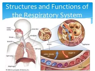
Bronchial Tree • Functions of the respiratory tubes and alveoli • Branches serve as passageways for air, filter incoming air, and distribute air evenly to alveoli in all parts of the lungs • Alveoli provide a large surface area for gas exchange • During gas exchange: • Oxygen diffuses through the alveolar walls and enters blood in nearby capillaries • Carbon dioxide diffuses from the blood through alveolar walls and enters the alveoli
Lungs • Suspended in the thoracic cavity by a bronchus and various large blood vessels • These tubular structures enter each lung on the medial surface through a region called the hilum • Covered by the visceral pleura which fold back to become the parietal pleura at the hilum • The visceral pleura and parietal pleura essentially touch each other and are separated only by a thin film of serous fluid (located in the pleural cavity) that serves as lubrication to reduce friction during breathing and helps to hold the pleural membranes together
Lungs • Right lung is larger than the left lung • The right lung is divided by fissures into the superior, middle, and inferior lobes • The left lung is divided by fissures into the superior and inferior lobes
Lungs • Lobes • Each lobe is supplied by a lobar bronchus, has connections to blood and lymphatic vessels, and is enclosed by connective tissues • Each lobe is further divided by connective tissues into lobules • Each lobule contains terminal bronchioles (along with their respective alveolar ducts, alveolar sacs, alveoli, nerves, blood vessels, and lymphatic vessels)