Download
1 / 16
220 likes | 510 Views
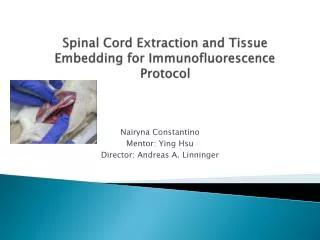
Spinal Cord Extraction and Tissue Embedding for Immunofluorescence P rotocol. Nairyna Constantino Mentor: Ying Hsu Director: Andreas A. Linninger. Research Update . Lumbar puncture practice Extraction of Spinal Cord Tissue embedding protocol

E N D
Spinal Cord Extraction and Tissue Embedding for Immunofluorescence Protocol NairynaConstantino Mentor: Ying Hsu Director: Andreas A. Linninger
Research Update • Lumbar puncture practice • Extraction of Spinal Cord • Tissue embedding protocol • Ordering materials for tissue embedding experiment • Performing experiments
Lumbar Puncture • Lumbar Puncture: -A procedure that collects the cerebrospinal fluid (CSF) -CSF: an important role in homeostatic, hormonal, and signaling functions -The fluid is found in the spaces around the brain and spinal cord -A patient lies in a curled position on a table. -A spinal needle is inserted into the lower part of the spinal column to remove cerebrospinal fluid (CSF, shown in blue).
Lumbar Puncture in rats Performing extraction of cerebrospinal fluid (CSF) Opening up the rat to get a better view of the spinal cord anatomy -Note: Live rat injection window will be smaller than 2 inches
Complications with extraction of CSF • Difficult to extract since: • 50-70 μL of CSF • Weight of rat spinal cord = 0.7 g • Spinal Cord Measurements
Spinal Cord Dimensions -Imaging of the rat spinal cord -Other scholars have observed as well how small the spinal cord is -Difficulty for lumbar practice -The spinal cord of a rat was examined -Diameter of spinal cord is approximately 2-3 mm -Difficulty for lumbar practice
Spinal cord in Cervical regionsof the rat -The spinal cord in the cervical regions as shown is approximately 2mm-3mm
Spinal Cord Extraction • Scissors are inserted between iliac crest of spinal cord and a cut is made adjacent to the cord • This is done on both sides such that the cord is freed along its lateral aspects from the body. • A second cut is then made transversely at a point 3 cm caudal to the last rib and at a point as rostral as the level of cord that is desired. • To remove the spinal cord from either block, the wooden dowel from a cotton swab isthen inserted caudal to rostral through the vertebral canal • Such an insertion forces the spinal cord out the other end.
Tissue embedding protocol • Method: Cut tissue to 2 mm in thickness(length/width) Place the tissue in the cassette Tissue labeled cassettes fixed in formalin solution (24 hours)
Protocol continued Embed the tissue in paraffin Remove tissue from cassette. Place paraffin into mold and press tissue against mold De-fat the tissue twice with xylene at room temperature Melt the paraffin by heating it in a heat proof vessel to 58-60 celsius Invert the tissue cassette over the mold. And pour paraffin on top of the cassette. Once paraffin hardens remove the block stir the cassette in paraffin Dehydration of tissue
Aquaporin channels Aquaporin 1 -Structure Å=1×10⁻¹⁰ m or 100 pm Tetramer -4 monomers Each monomer provides independent water pore Six transmembrane helices are packed to form the trapezoid structure
Aquaporin 1 Dimensions Dimensions Format 1 Dimensions Format 2 60 Å= 6 nm 40 Å= 4 nm
AQP1 in ribbon format Monomer -Six transmembrane helices Pore is shown in blue See the three parts to a monomer: Extracellular Pore (selectivity filter) Cytoplasmic vestibule
Three parts of a monomer Dimensions: Extracellular -diameter = 15 Å (1.5 nm) Pore (sensitivity filter) -Long=20 Å ( 2 nm) -Down this pore there is a constriction section. -After passing the constriction section the pore has a diameter of 4 Å (0.4 nm) Cytoplasmic vestibule -Mouth of 15 Å (1.5 nm) wide
References • http://www.pnas.org/content/94/10/5034/F2.expansion.htm5034/F2.expansion.htm • http://www.cancer.gov/cancertopics/pdq/treatment/adultAML/Patient/page2/Print • Anatomy of the Laboratory Rat. Rudolf Hebel & M.W. Stromberg • http://wwww.cti.northwestern.edu/sa/scan • http://www.christopherreeve.orge/site/c.dd|Fig/b.4819993/k.4888/Spinal_Cord_Atlas_Rat_Spinal_Cord.htm