Download

1 / 87
870 likes | 1.34k Views
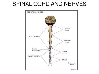
Spinal Cord and Spinal Nerves. Spinal Cord. Enclosed in the vertebral canal, extends from the foramen magnum of the skull to the first or second lumbar vertebra where it terminates in the cone shaped conus medullaris. Spinal Cord. Spinal meninges; Dura Mater – outer Arachnoid Mater – middle

E N D
Spinal Cord • Enclosed in the vertebral canal, extends from the foramen magnum of the skull to the first or second lumbar vertebra where it terminates in the cone shaped conus medullaris
Spinal Cord • Spinal meninges; • Dura Mater – outer • Arachnoid Mater – middle • Pia Mater – inner
Spinal Cord • Dura Mater – composed of dense, irregular connective tissue
Spinal Cord • Arachnoid Mater – It is an avascular covering with a spider’s web arrangement of delicate collagen fibers and some elastic fibers
Spinal Cord • Pia Mater – A vascular and thin transparent connective tissue layer composed of interlacing bundles of collagen fibers and some fine elastic fibers
Spinal Cord • Subarachnoid space – between the arachnoid mater and pia mater which contains cerebrospinal fluid
Spinal Cord • The dura mater and arachnoid meningeal coverings extend beyond the conus medullaris, approximately to the level of S2
Spinal Cord • Filium terminale – a fibrous extension of the pia mater, extends farther and attaches to the posterior coccyx
Spinal Cord • 31 pairs of spinal nerves, which exit via the intervertebral foramina
Spinal Cord • Since the spinal cord doesn’t extend to the end of the vertebral column, the spinal nerves emerging from the inferior end must travel through the vertebral canal until reaching the appropriate intervertebral foramina
Spinal Cord • This collection of spinal nerves are called the cauda equina
Spinal Cord / Gray Matter • Looks like an H
Spinal Cord / Gray Matter • Posterior or dorsal horns – posterior projections
Spinal Cord / Gray Matter • Anterior or ventral horns – anterior projections that contain cell bodies of motor neurons
Spinal Cord / Gray Matter • Lateral Horns – In the thoracic and lumbar regions there is a lateral outpocketing of gray matter
Spinal Cord / Gray Matter • Gray commissure – central area of gray matter
Spinal Cord / Gray Matter • Dorsal root – Sensory fibers enter the cord here
Spinal Cord / Gray Matter • Dorsal Root Ganglia – Cell bodies of sensory neurons located here
Spinal Cord / Gray Matter • Ventral Roots – Motor neurons leave the cord here
Spinal Cord / Gray Matter • Spinal nerves – are formed from the fusion of the dorsal and ventral roots
Spinal Cord / Gray Matter Spinal Nerves; 8 pairs of cervical nerves 12 pairs of thoracic nerves 5 pairs of lumbar nerves 5 pairs of sacral nerves 1 pair of coccygeal nerves
Spinal Cord / White Matter • The anterior median fissure and the posterior median sulcus divide the spinal cord into R. and L. sides
Spinal Cord / White Matter • White matter is divided into columns; Posterior Funiculus, Anterior Funiculus, and Lateral Funiculus
Spinal Cord / White Matter • Each column contains distinct bundles of nerve axons called tracts
Spinal Cord / White Matter Two Types of Tracts 1. Sensory (ascending) tracts – conduct nerve impulses toward the brain
Spinal Cord / White Matter 2. Motor (descending) tracts – conduct impulses down the cord
Connective Tissue Coverings of Spinal Nerves • A fiber is a single axon within an endoneurium
Connective Tissue Coverings of Spinal Nerves • A fascicle is a bundle of fibers within a perineurium
Connective Tissue Coverings of Spinal Nerves • A nerve is a bundle of fascicles within an epineurium
Spinal Nerves and Nerve Plexus • Each nerve divides into dorsal and ventral rami
Spinal Nerves and Nerve Plexus • Rami contains both motor and sensory rami
Spinal Nerves and Nerve Plexus • Dorsal rami – serve the skin and musculture of the posterior body trunk at their approximate level of emergence
Spinal Nerves and Nerve Plexus • Ventral rami of spinal nerves T2 –T12 – pass anteriorly to supply the muscles of intercostal spaces, and the skin and muscles of the anterior and lateral trunk
Spinal Nerves and Nerve Plexus • Ventral rami of all other nerves – form complex networks of nerves called plexuses