Download
1 / 35
400 likes | 625 Views
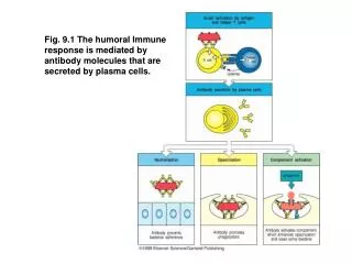
Fig. 9.1 The humoral Immune response is mediated by antibody molecules that are secreted by plasma cells. Fig. 9.2 A second signal is required for B-cell activation by either thymus-dependent or thymus-independent antigens.

E N D
Fig. 9.1 The humoral Immune response is mediated by antibody molecules that are secreted by plasma cells.
Fig. 9.2 A second signal is required for B-cell activation by either thymus-dependent or thymus-independent antigens
Fig. 9.3 B cells and helper T cells must recognize epitopes of the same molecular complex in order to interact.
Protein antigens attached to polysaccharide antigens allow T cells to help polysacharide-specific B cells.
Fig. 9.5 Armed helper T cells stimulate the proliferation and then the differentiation of antigen-binding B cells.
Fig. 9.6 When an armed helper T cell encounters an antigen-binding B cell, it becomes polarized and secretes IL-4 and other cytokines at the point of cell-cell contact.
Fig. 9.7 Different cytokines induce switching to different isotypes.
Fig. 9.8 Isotype switching is preceded by transcriptional activation of heavy-chain C-region genes.
Fig. 9.9 Antigen-binding cells are trapped in the T-cell zone.
Fig. 9.10 Plasma cells secrete antibody at a high rate but can no longer respond to antigen or helper T cells.
Fig. 9.11 Activated B cells form germinal centers in lymphoid follicles.
Fig. 9.12 Germinal centers are formed when activated B cells enter lymphoid follicles.
Fig. 9.13 After T-cell-dependent activation , B cells undergo rounds of mutation and selection for higher-affinity memory B cells and antibody secreted from plasma cells.
Fig. 9.14 Immune complexes bind to the surface of follicular dendritic cells.
Fig. 9.15 Immune complexes bound to follicular dendritic cells form iccosomes, which are released and can be taken up by B cells in the germinal center.
Fig. 9.16 Thymus-independent type 1 antigens (Tl-1 antigens) are polyclonal B-cell activators at high concentrations, whereas at low concentrations they induce an antigen-specific antibody response.
Fig. 9.17 B-cell activation by thymus-independent type 2 antigens (Tl-2 antigens) requires, or is greatly enhanced by, cytokines.
Fig. 9.18 Properties of different classes of antigen that elicit antibody responses.
Fig. 9.19 Each human immunoglobulin isotype has specialized functions and a unique distribution.
Fig. 9.20 Transcytosis of lgA antibody across epithelia is mediated by the poly-lg receptor, a specialized transport protein.
Fig. 9.22 Immunoglobulin isotypes are selectively distributed in the body.
Fig. 9.23 Many common diseases are caused by bacterial toxins.
Fig. 9.24 Neutralization of toxins by lgG antibodies protects cells from their damaging action.
Fig. 9.25 Viral infection of cells cqan be blocked by neutralizing antibodies.
Fig. 9.26 Antibodies can prevent attachment of bacteria to cell surfaces.
Fig. 9.28 The classical pathway of complement activation is initiated by binding of C1q to antibody on a surface such as a bacterial surface.
9.29 Erythrocyte CR1 helps to clear immune complexes from the circulation.
Fig. 9.30 Distinct receptors for the Fc region of the differentimmunoglobulin isotypes are expressed on different accessory cells.
Fig. 9.31 Bound antibody is distinguishable from free immunoglobulin by its state of aggregation.
Fig. 9.32 Fc and complement receptors on phagocytes trigger the uptake and degradation of antibody-coated bacteria.
Fig. 9.33 Eosinophlis attacking a schistosome larva in the presence of serum from an infected patient.
Fig. 9.34 Antibody –coatedtarget cells can be killed by NK cells in antibody-dependent cell-mediated cytoxicity.
Fig. 9.35 lgE antibody cross-linking on mast-cell surfaces leads to a rapid release of inflammatory mediators.