Effects of UVB Irradiation and DIC on Skin Erythema and Wrinkle Formation in Hairless Mice
This study investigates the minimal erythema dose (MED) in UVB-irradiated hairless mice, identifying the MED at 120 mJ/cm². We explore the anti-wrinkle effects of DIC, an elastase inhibitor, and its inability to prevent collagen fiber degradation induced by UVB. Additionally, we analyze the impact of KPA and chymostatin on fibronectin fragmentation and the conversion of proMMP-1 to active MMP-1. Findings reveal the limitations of DIC in promoting skin health post UVB exposure, highlighting the complex interactions involved in skin aging and damage repair.

Effects of UVB Irradiation and DIC on Skin Erythema and Wrinkle Formation in Hairless Mice
E N D
Presentation Transcript
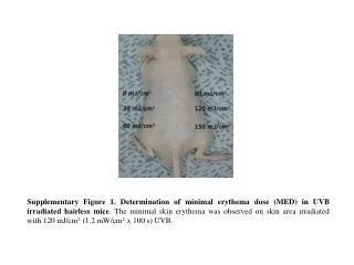
Supplementary Figure 1. Determination of minimal erythema dose (MED) in UVB irradiated hairless mice. The minimal skin erythema was observed on skin area irradiated with 120 mJ/cm2 (1.2 mW/cm2x 100 s) UVB.
UVB (13 W) (a) Non-UV Vehicle DIC (0.01%) (b) (c) Supplementary Figure 2. The anti-wrinkle effects of DIC, an elastase inhibitor. DIC did not attenuate wrinkle formation and degradation of collagen fibers by UVB (a) Replica photography (b) Massons’s trichrome staining for collagen fibers (c) Verhoeff staining for elastin fibers. Magnification: x 200. Bar: 200 ㎛.
(b) (a) KPA(µM) Chymostatin (µg/ml) DIC (µM) - 0.8 0.16 - 10 4 - 0.8 0.16 - + + + + + + - + + + CG (1mU/ml) Intact Fn 191 97 64 51 39 28 19 (kDa) LC Fn-fr’s (c) (d) Chymostatin KPA DIC (µg/ml) (µM) (µM) 10 4 2 0.8 0.16 0.0032 0.8 0.16 - - + + + + + + + + + + + CG (1mU/ml) Pro MMP-1 Active MMP-1 Supplementary Figure 3. The inhibitory effects of KPA, chymostatin and DIC on fibronectin fragmentation and conversion of proMMP-1 to active MMP-1 in cathepsin G (CG) treated NHFs. The fragmentation of Fn was analyzed in presence of KPA, chymostatin and DIC by western blot using anti-fibronectin Ab. (a, c) Specific inhibitors of cathepsin G like KPA and chymostatin prevented Fn degradation and conversion of proMMP-1 to active MMP-1 by cathepsin G (18). (b, d) DIC, an elastase specific inhibitor, did not prevented Fn fragmentation and conversion of proMMP-1 to active MMP-1 by cathepsin G.
Con CG (mU) - 1 5 10 Con Elastase (mU) - 1 5 Pro MMP-1 Active MMP-1 Supplementary Figure 4. Conversion of proMMP-1 to active MMP-1 by cathepsin G (CG) or elastase treatment in NHFs. Cathepsin G promoted the conversion of proMMP-1 to active MMP-1, but elastase did not.
(a) (b) Supplementary Figure 5. Pictures of 3D dermal equivalent (3D DE). 3D DE contains collagen, fibronectin and normal human fibroblasts inDMEM medium. It changes from liquid state to gel state after incubation at 37℃ for 2 hour. (a) 3D DE in 24 well plates and (b) gel state 3D DE out 24 well plates.
(a) Non UV (b) UVB+Vehicle (c) UVB+ RA (0.01%) (d) UVB+ KPA (0.025%) Supplementary Figure 6. Verhoeff staining of elastic fibers after KPA treatment. (a) Non UV control (b) UVB-irradiated vehicle treated group (c) UVB-irradiated retinoic acid treated group (d) UVB-irradiated KPA treated group. Magnification: x 200. Bar: 200 ㎛