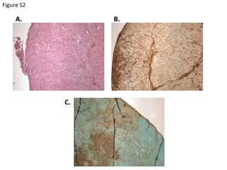
Figure 15.25a
Middle ear. Internal ear (labyrinth). External ear. Auricle (pinna). Helix. Lobule. External acoustic meatus. Tympanic membrane. Pharyngotympanic (auditory) tube. (a) The three regions of the ear. Figure 15.25a. External Ear. Auricle ( pinna ) composed of: Helix (rim)
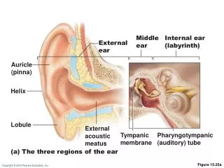

Figure 15.25a
E N D
Presentation Transcript
Middle ear Internal ear (labyrinth) External ear Auricle (pinna) Helix Lobule External acoustic meatus Tympanic membrane Pharyngotympanic (auditory) tube (a) The three regions of the ear Figure 15.25a
External Ear • Auricle (pinna) composed of: • Helix (rim) • Lobule (earlobe) • External acoustic meatus (auditory canal) • Ceruminousglands
External Ear • Tympanic memb(eardrum) • Between external and middle ear • Connective tissue membrane vibrates in response to sound • Transfers sound energy to bones of middle ear
Middle Ear • Air-filled, mucosa-lined cavity in temporal bone • Pharyngotympanic (auditory) tube—connects middle ear to nasopharynx • Equalizes pressure with external air pressure
Oval window (deep to stapes) Semicircular canals Entrance to mastoid antrum in the epitympanic recess Malleus (hammer) Vestibule Incu (anvil) Auditory ossicles Vestibular nerve Stapes (stirrup) Cochlear nerve Tympanic membrane Cochlea Round window Pharyngotympanic (auditory) tube (b) Middle and internal ear Figure 15.25b
Ear Ossicles • Three small bones in tympanic cavity: malleus, incus, and stapes • Suspended by ligaments and joined by synovial joints • Transmit vibration of eardrum to oval window • Tensor tympani and stapedius muscles contract reflexively in response to loud sounds to prevent damage to hearing receptors
Epitympanic recess Malleus Incus Superior Lateral Anterior View Pharyngotym- panic tube Tensor tympani muscle Tympanic membrane (medial view) Stapes Stapedius muscle Figure 15.26
Internal Ear • Bony labyrinth • Channels in temporal bone • Three parts: vestibule, semicircular canals, and cochlea • Filled with perilymph • Membranous sacs within bony labyrinth • Filled with K+ rich endolymph
Superior vestibular ganglion Inferior vestibular ganglion Temporal bone Semicircular ducts in semicircular canals Facial nerve Vestibular nerve Anterior Posterior Lateral Cochlear nerve Cristae ampullares in the membranous ampullae Maculae Spiral organ (of Corti) Utricle in vestibule Cochlear duct in cochlea Saccule in vestibule Stapes in oval window Round window Figure 15.27
Vestibule • Two membranous sacs • Saccule- continuous with cochlear duct • Utricle - continuous with semicircular canals • House equilibrium receptor regions (maculae) • Respond to gravity and changes in position of head
The Cochlea • Coils around a bony pillar (modiolus) • Contains cochlear duct, which houses spiral organ (of Corti) and ends at cochlear apex
The Cochlea • Cochlea is divided into 3 chambers • Scalavestibuli—abuts oval window, contains perilymph • Scala media (cochlear duct)—contains endolymph • Scala tympani—terminates at round window; contains perilymph • Scalae tympani and vestibuli continuous with each other at helicotrema (apex)
The Cochlea • “Roof” of cochlear duct is vestibular membrane • “Floor” of cochlear duct is: • Bony spiral lamina • Basilar membrane, which supports organ of Corti • Cochlear branch of nerve VIII runs from organ of Corti to brain
Modiolus Cochlear nerve, division of the vestibulocochlear nerve (VIII) Spiral ganglion Osseous spiral lamina Vestibular membrane Cochlear duct (scala media) Helicotrema (a) Figure 15.28a
Vestibular membrane Osseous spiral lamina Tectorial membrane Spiral ganglion Scala vestibuli (contains perilymph) Cochlear duct (scala media; contains endolymph) Stria vascularis Spiral organ (of Corti) Scala tympani (contains perilymph) Basilar membrane (b) Figure 15.28b
Tectorial membrane Inner hair cell Hairs (stereocilia) Afferent nerve fibers Outer hair cells Supporting cells Fibers of cochlear nerve Basilar membrane (c) Figure 15.28c
Inner hair cell Outer hair cell (d) Figure 15.28d