Download
1 / 1

E N D
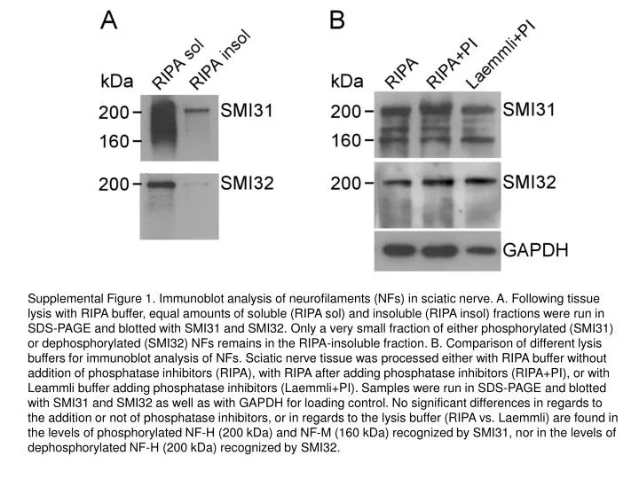
Supplemental Figure 1. Immunoblot analysis of neurofilaments (NFs) in sciatic nerve. A. Following tissue lysis with RIPA buffer, equal amounts of soluble (RIPA sol) and insoluble (RIPA insol) fractions were run in SDS-PAGE and blotted with SMI31 and SMI32. Only a very small fraction of either phosphorylated (SMI31) or dephosphorylated (SMI32) NFs remains in the RIPA-insoluble fraction. B. Comparison of different lysis buffers for immunoblot analysis of NFs. Sciatic nerve tissue was processed either with RIPA buffer without addition of phosphatase inhibitors (RIPA), with RIPA after adding phosphatase inhibitors (RIPA+PI), or with Leammli buffer adding phosphatase inhibitors (Laemmli+PI). Samples were run in SDS-PAGE and blotted with SMI31 and SMI32 as well as with GAPDH for loading control. No significant differences in regards to the addition or not of phosphatase inhibitors, or in regards to the lysis buffer (RIPA vs. Laemmli) are found in the levels of phosphorylated NF-H (200 kDa) and NF-M (160 kDa) recognized by SMI31, nor in the levels of dephosphorylated NF-H (200 kDa) recognized by SMI32.