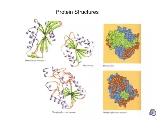
Protein Structures
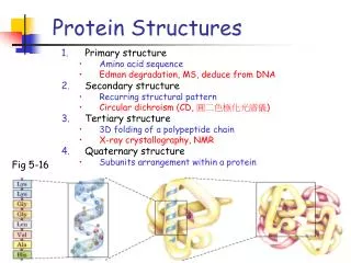
Protein Structures. Primary structure Amino acid sequence Edman degradation, MS, deduce from DNA Secondary structure Recurring structural pattern Circular dichroism (CD, 圓二色極化光譜儀 ) Tertiary structure 3D folding of a polypeptide chain X-ray crystallography, NMR Quaternary structure

Protein Structures
E N D
Presentation Transcript
Protein Structures • Primary structure • Amino acid sequence • Edman degradation, MS, deduce from DNA • Secondary structure • Recurring structural pattern • Circular dichroism (CD, 圓二色極化光譜儀) • Tertiary structure • 3D folding of a polypeptide chain • X-ray crystallography, NMR • Quaternary structure • Subunits arrangement within a protein Fig 5-16
The 3-D structure of proteins • Protein conformation in space • Including long-range interactions • Determined by: • Primary (and secondary) structures • Interactions among R groups • Disulfide bond and weak interactions
Unfolded (denatured) High degree of conformational entropy H-bond of polypeptide with solvent (H2O) Folded (native) Lowest free energy Stabilized by disulfide bond (covalent) and weak (non-covalent) interactions: Weak interactions Van der Waals interaction H-bond Hydrophobic Ionic Protein stability In general, the protein conformation with lowest free energy is the one with the max. no. of weak interactions.
OC-NH is shorter Coplanar peptide group Trans configuration (O vs. H) Electrons resonance (partial sharing) between the carbonyl O and the amide N. (electric dipole) OC-NH can not rotate Limited rotation for Ca-C (, psi) and N-Ca (, phi) Peptide bond
Collagen triple helix Anti-parallel b-sheet Parallel b-sheet Right-handed a-helix Protein secondary structure • Local conformation, regular backbone pattern • Restricted and in 2o structures • Determined by primary structure • a-helix (e.g. a-keratin in hair) • b-sheet (e.g. silk fibroin – layers of b-sheets) • b-turn Ramachandran plots
1 turn R-- -- R Box 6-1 a-helix A right-handed a-helix: • 3.6 a.a. per turn • 5.4 Å (1 Å = 0.1 nm) per turn • R groups extended outwardperpendicular to the helical axis • H-bonding between adjacent turns • H-bond between the -CO of residue (i) and the -NH of residue (i+3). • 2 H-bonds per residue • 3 or 4 H-bonds per turn • Provide stability
- O O H3N+ C O- H3N+ C O- CH CH2 CH CH2 CH2 H + a-helix constraints • Electrostatic interactions of Ri and Ri+1 • Size of the R group • Interactions between Ri and Ri+3 or Ri+4 • Pro and Gly • End residues (electric dipole)
- Fig 6-2a + Electric dipole of an a-helix • Peptide bond dipole • Helix dipole • End residues and helix stability Fig 6-6
parallel antiparallel b-conformation • Zigzag, extended protein chain, with the R groups alternating above and below the backbone. • Side by side b-conformation b-sheet • H-bonds between adjacent peptide chain (backbone). • Parallel or antiparallel orientations • Silk fibroin – layers of b-sheets
3 (Gly) 3 4 4 Fig 6-8a 2 2 1 1 b-turn • A 180o turn involving 4 a.a. • H-bond between -CO of the 1st a.a. and the -NH of the 4th a.a. • Common a.a. • Gly (small and flexible, type II b-turn) • Pro (peptide bonds involving the imino N in cis configuration)
Occurrence in 2o structure • Relative probability of a.a. Fig 6-10
Circular Dichroism Spectroscopy • Determine the content of 2o structure of a protein http://www-structure.llnl.gov/cd/cdtutorial.htm
Membrane proteins Lehninger 4th ed. • Membrane spanning protein (hydropathy plot, p. 377) • a helix type channels (helical wheel diagram, p. 393) • b barrel porins (p. 378)
Classification (p. 170) • Fibrous proteins (e.g. Table 6-1) • Long strands or sheets • Consist of a single type of 2o structure • Function in structure, support, protection • a-keratin, collagen • Globular proteins (e.g. Table 6-2) • Spherical or globular shape • Contain several types of 2o structure • Function in regulation • Myoglobin, hemoglobin
Structure of hair a-keratin: hair, wool, nails, claws, quills, horns, hooves, and the outer layer of skin Fig 6-11, p. 171 Monomer Dimmer
collagen tropocollagen Collagen • Tendons, bone, cartilage, skin, and cornea • Primary sequence: • Gly-X-Pro (HyPro) • Repeating tripeptide unit • Structure • Monomer (a chain) • Left-handed helix, 3 a.a. per turn • Trimer: coiled-coil (tensile strength). • Stabilized by H-bond • Crosslink between triple helixes • Genetic defect: • Osteogenesis imperfecta • Abnormal bone formation in babies • Ehlers-Danlos syndrome • Loose joint
More on Collagen … Harper’s 26th, p. 38-39. • Procollagen (a larger precursor polypeptide) • Post-translational modification • Pro, Lys Hydroxyl Pro, Lys (cofactor = ascorbic acid) • Provide H-bond that stablizes the mature protein • Scurvy: a dietary deficiency of Vit C • Central portion triple helix (procollagen collagen) • The N-, and C-terminal portions are removed • Certain Lys are modified by lysyl oxidase (a copper-containing protein) • Crosslink between polypeptides increased strength and rigidity. • Menke’s syndrome: a dietary deficiency of the copper
Denature and unfolding • Loss of function due the structural disruption • Cooperative process • Denatured conformation: random but partially folded • No covalent bonds in the polypeptide are broken !! • Denaturing agent • Heat (H-bond) • Extreme pH (change ionic interaction) • Miscible organic solvent (hydrophobic interactions) • Alcohol, acetone • Certain solutes (hydrophobic interactions) • Urea, guanidino hydrochloride (Gdn HCl), detergent No function Fully functional
The prion disease • Spongiform encephalopathies • Disease caused by a protein (prion) • Proteinaceous infectious particle • Related diseases: • Mad cow disease • Kuru • Creutzfeldt-Jakob disease (human) • Scrapie (sheep) • Misfolded prion PrPC (normal) PrPSC (infectious)
Protein Function Myoglobin and Hemoglobin
O2 binding to Heme • Heme = organic ring (porphyrin) + Fe2+ • Free heme Fe2+ (binds O2) vs. Fe3+ (does not bind) • O2 rich blood (bright red) vs. O2 depleted blood (dark purple) • CO, NO binds with higher affinity than O2
[PL] Ka: association constant (M-1) Ka = [P] [L] [PL] Ka [L] = [P] Binding sites occupied [PL] = = Total binding sites [L] [L] [PL] + [P] = = Kd: dissociation constant (M) [L] + 1/Ka [L] + Kd Protein-ligand interaction p. 207 • P + L PL
[L] = [L] + Kd Ligand binding and Kd • When [L] = Kd, 50% ligand-binding sites are occupied • Kd: dissociation constant • Kd = [L] at half-saturation • Affinity , Kd Hyperbola Fig 7-4a
tissues lungs [L] = [L] + Kd pO2 = pO2 + P50 O2 binding of Mb • O2 binds tightly to Mb • Good for O2 storage • Not good for O2 transport 1 atm = 105 Pa = 100 kPa pO2, air = 20 kPa 0.26 kPa Fig 7-4b
Steric hindrance • Distal His, (His64 of Mb) • Molecular motion (breathing) • O2 in/out buried cavity Structure affects Kd Kd for O2 Kd for CO • Free heme 1x 1/20,000x • Heme in Mb 1x 1/200x
O2 transport Found in erythrocyte Hb = tetramer 4 x (polypeptide chain + heme) Hb m.w. = 64.5 KDa Interactions between subunits (tetramer) O2 storage In muscle tissue Mb = monomer 1 polypeptide chain (153 a.a.) + 1 heme Mb m.w. = 16.7 kDa Mb vs. Hb Sequence vs. structure homology Fig 7-3 Fig 7-10
Hb has 2 conformations T state R state -O2 structure stable unstable +O2 unstable stable Kd (O2) large small • O2 binding to T triggers a conformational change to R Fig 7-10
= 0.96 = 0.64 Hb–O2 binding curve • A sigmoid (S-shape) binding curve • Permit highly sensitive response to small change in pO2 or [ L] Fig 7-12
O2 binding to Hb • Cooperativity • One subunit binding of O2 affects Kd of the adjacent subunits • 4 x (subunit + O2) • 1st O2 binds Hb (T) weakly, initiate T R • 2ndO2 binds Hb (TR) with higher affinity • 3rd O2 binds Hb (TR) with even higher affinity • 4th O2 binds Hb (R) with highest affinity • S-shaped (sigmoid) binding curve – multimer only • Allosteric protein • Homotropic: modulator = ligand (substrate) • e.g. O2, CO • Heterotropic: modulator ligand (substrate) • e.g. H+, CO2, BPG
[PLn] Ka = [P] [L]n Binding sites occupied [L]n = = Total binding sites [L]n + Kd [L]n = 1 - Kd log n log [L] – log Kd = 1 - Quantification • P + n L PLn Slope = n (Hill coefficient) n > 1, + Coop. n = 1, no Coop. n < 1, - Coop. log 1 - Y = ax - b log [L] Hill equation
Hill plot of Mb vs. Hb • Mb: nH = 1 • Hb: nH = 3 Fig 7-13
Hb also transports H+ and CO2 • Bohr effect • pH and CO2 modulate the affinity of Hb for O2 • Hb binds O2 and (H+ or CO2) with inverse affinity • Hb binds O2, H+, and CO2 at different sites • Tissues: pH and CO2, O2 affinity , Hb release O2 • Lungs: pH and CO2, O2 affinity , Hb binds more O2 In lung In tissue
BPG (2,3-bisphosphoglycerate) • BPG binds to a.a. in the cavity between b subunitsin Hb (T state) • BPG stabilize T state O2 affinity • [BPG] at sea level vs. high altitude • Fetal Hb – needs to have a higher O2 affinity than mother’s Hb • Fetal Hb : a2g2 • [BPG] , after storage, transfusion… • People suffering from hypoxia, [BPG]↑…
CO intoxication (Box 5-1) • CO has a higher affinity for Hb • Smoker has higher level of COHb (3~15%) vs. < 1% • Binding of CO to Hb increase the O2 affinity of Hb • O2 transport become less efficient (Fig 2) • Suspected CO intoxication • Rapid evacuation • Administer 100% O2 Lehninger 4th ed.
Sickle-cell anemia • Homozygous allele for the b subunit gene • Hb A (Glu6) vs. Hb S (Val6) on b subunits surface • “Sticky” hydrophobic contacts • deoxyHb S: insoluble and form aggregates • Heterozygous: malaria resistance • Anemia or Malaria ? HbA